Total Pageviews

Translate

Followers

Sunday, February 5, 2023

Big Toe Joint Pain (Sesamoid Injury): One Example on How to Work Up the Patient using 14 Point Assessment Summary


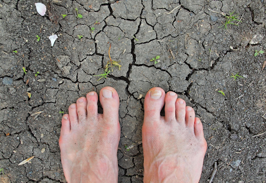
 Patient #17: Big Toe Joint Pain 

                                                 . 

History and Chief Complaint

  • Long distance runner presents with a 6 month history of progressively worsening pain at the ball of his right foot
  • The pain had be smoldering for awhile, but got much worse when he attempted stair running at a local stadium
  • Over the last 2 months, he could not walk well, so he got an appointment with the local podiatrist
  • X Rays were negative by history, and he was placed in a removable boot for several months
  • The pain was not any better in the boot, painful with each step, and a roommate gave him some old crutches stored at his mom’s house
  • The big toe joint was swollen and painful to move
  • Follow up appointment with the podiatrist an MRI was ordered. 
  • The doctor said that he had a stress fracture in the medial sesamoid and that time or surgery would heal.
  • He came to me for a second opinion only

Gait Evaluation

  • Very limited due to the boot and need for crutches
  • When asked, the patient stated he had been labelled a pronator
  • Running shoes were not present, but minimalist in nature
  • Gait evaluation for walking and running would have to be delayed (it can be months before I watch a runner actually run due to situations like this)

Physical Examination

  • Swollen first metatarsal phalangeal joint
  • Palpable pain plantar only on both sesamoids, perhaps the medial more
  • Plantar pain on both maximum dorsiflexion and plantarflexion of the joint
  • Plantar pain on contraction of the flexor hallucis longus against resistance
  • Good range of motion of the joint however, although 10 degrees less overall motion than uninvolved side
  • Probable negative lachman (swelling present can make the test unreliable)
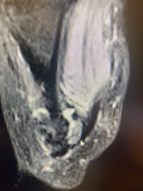
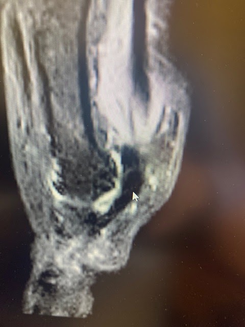
  • MRI showed no apparent fracture but bone edema in the medial sesamoid and surrounding tissue

Cursory Biomechanical Examination  and Asymmetry Noted

  • Rigid Pes Cavus Foot Type
  • Inverted Heel RCSP
  • Mild Tight Achilles Tendons
  • Plantar Flexed First Ray Right Worse than Left (perhaps only due to swelling)
  • Everted Forefoot Deformity Left greater than right

Tentative Working Diagnosis 

  • Medial Sesamoid Stress Fracture (stress fractures may not be seen even on MRI)

Common Differential Diagnosis (2ndary Working Diagnosis)

  • Sesamoiditis with Bone Edema

Occam’s Razor and Rule of 3

  • Simplest Solution after 6 months of pain and on crutches and a boot is surgical removal (no one would think that wrong)
  • Rule of 3 looks for ways to rehabilitate, although can be also used post operatively to have a better outcome.
  • The 3 forces that lead to stress in the sesamoids commonly are: tight achilles tendons that must be stretched out, pes cavus feet with high metatarsal declination angles that need to have the re-balanced, and plantar flexed first rays that must be off loaded

What Phase of Rehabilitation?

  • Immobilization (but non-weight bearing with crutches can intensify the swelling accumulation and make the patient feel worse than they actually are)

Should We Image?

  • X Rays and MRI already done
  • I always look at the first MRI as just that “The First”
  • I will get another MRI in 6 months to see how the healing is going

First Decision: How to Reduce Pain 0-2

  • Minimal to No Crutches as non weight bearing increases swelling to a localized area like this
  • Build an accommodation of at least ¼ inch adhesive felt inside of the removable boot (normally up to ½ inch)

Second Decision: Inflammation Concerns

  • No NSAIDs due to bone problem
  • Ice Packs or Soaks 3 times a day 
  • Begin Contrast Bathing each evening to get rid of the bone edema

Third Decision: Any Nerve Component?

  • Assume that nerve hyper-sensitivity begins 3 months after a problem like this.
  • Start treating with ice for only 5 minutes, warm compresses, non painful massage, topical gels or patches (like Neuro Eze lotion or Lidoderm Patches)

Fourth Decision: Initial Mechanical Changes

  • Get the boot comfortable so that we know we have a healing environment (make an internal float)
  • Order a 9 month course of Exogen Bone Stimulator
  • Begin designing or ordering off weighting pads like Dr. Jill's Dancer’s Pads of a ¼ inch thick

     This particular patient did well with conservative treatment over the next several years and was back running. The mechanical list from Chapter 6 (Book 2 of Practical Biomechanics for the Podiatrist) is so long due to the long period of experimentation needed for a patient like this. I have starred all the ones used for this particular patient as we moved him through his rehabilitation. 


Common Mechanical Changes at the First Metatarsal Phalangeal Joint

  1. Spica Taping*
  2. Bunion Taping
  3. Toe Separators
  4. Dancer’s Padding*
  5. Cluffy wedges*
  6. Morton’s extensions
  7. Orthotic Devices for Weight Shift with no extrinsic post*
  8. Orthotic Devices of Stability only with no extrinsic post 
  9. No Heel Lift*
  10. Zero Drop Shoes*
  11. Rocker Shoes*
  12. Bike Shoes with Embedded Cleats
  13. Cam Walkers or Removable Boots*
  14. Stiff Shoes (including post operative shoes)*
  15. Flexible Shoes
  16. Forefoot Padding
  17. Skip Lacing*
  18. Deep Toe Box*
  19. Wide Toe Box
  20. Shoe Stretching
  21. Carbon Plate Full
  22. Carbon Plate Morton’s Extension
  23. Carbon Plate Dancer’s Modification
  24. Proximal Padding Dorsal or Medial
  25. Metatarsal Padding sub 2nd through 4th or 5th*
  26. Self Mobilization for Hallux Limitus
  27. Metatarsal Doming*
  28. Abductor Hallucis Strengthening
  29. Flexor and Extensor Hallucis Longus Tendon strengthening*
  30. Night Splints and Yoga Toes
  31. Correct Toes
  32. No Achilles Tightness*
  33. Avoid Excess Toe Bend*


     I saw this patient once a month for a year to progress the rehabilitation. He was in the boot for almost 3 months from the day I first saw him. While in the boot, he was working on bone health with  diet and bone stimulator, and he was working on the inflammation and nerve sensitivity 5 separate times each day. I should have at least got a Vit D blood level, as transient vitamin D deficiencies can lead to bone issues and prevent or slow down healing.The 2nd 3 months was still partial boot as we weaned him off the boot and into Hoka One One Shoes 1 size bigger and the widest one on the market. I needed room to build things for him. It was at 9 months along our rehab course when he had built up his pain free walking to 5 minutes, that he started the 10 level Walk Run Program. At one year from the start of seeing him, he ran a slow 10K in 73 minutes. In the shoes that he ran in were Hannafords (full length soft based plastazote custom inserts). I had made him a pair of Root Balanced plastic based orthoses due to the lateral column support I needed to get (high everted forefoot deformities) which worked best as scaled down dress orthotic devices. I had made him an Inverted Pair, but he was too laterally unstable as he began to walk and run. I also think the arch began too high in the Inverted pair, so the metatarsal declination pitch became too high. However, rehabilitation of these conditions, with all the modalities at our disposal is pretty awesome. 

Friday, February 3, 2023

Skip Lacing for Sore Spots


Skip Lacing to Avoid a Sore Area Top of Foot

          Today I worked with Dr Ajitha Nair, in Oakland California, on a patient with dorsal (top) foot pain. The ability to skip laces, but try to obtain as little loss in stability as possible, is crucial for these issues. Here two laces are kept by the holes, instead of criss crossing over the top of the foot. Once the area heals, the lacing may be returning to normal. We always give 2-3 weeks of feeling fine, before switching back.  

Thursday, February 2, 2023

Sesamoid MRIs: What is Going On? Is There Still Healing Occurring?

T2 images showing inflamed fibular sesamoid

T2 images showing normal tibial sesamoid
Greeting from San Francisco

T1 image showing intact stable but injured fibular sesamoid. The grey part of the bone indicates bone activity. 

Another T1 image of injured fibular sesamoid. Healing fine. No AVN or avascular necrosis

Great T1 image of both sesamoids. The injured fibular sesamoid has the re-moldeling area in the middle which looks more grey. No AVN or fragmentation.

     I have shown both T1 (normal bone white) and T2 (normal bone black) images. When a patient develops AVN, both T1 and T2 are black. Definitely not in this case. 

Monday, January 30, 2023

Great Videos on the Nerves of the Foot, Ankle and Calf

     Dealing with patients with nerve pain all the time, this is a great video that I could watch over and over again. I have watched it several times today, and learned something that I had forgotten about the deep peroneal nerve for instance. 


My next blog post will be a 10 question test from points made in these videos. And, the next blog post will be the answers. 

Friday, January 27, 2023

Tuesday, January 24, 2023

RED-S. Relative Energy Deficiency--Sport (Syndrome)

     This is a must read for any health care provider treating athletes! Thank you Dr. Karen Langone for pointing it out to me! 


https://www.childrenshospital.org/conditions/red-s

Progressive Collapsing Foot Deformity Classification by Dr Allen Jacobs


https://www.hmpgloballearningnetwork.com/site/podiatry/whats-name-understanding-progressive-collapsing-foot-deformity?hmpid=&utm_medium=email&utm_source=enewsletter&utm_content=1710811546

Patent Leather Shoe Stretching: This Article gives some Tips


  I had a patient yesterday with bunion irritation from her beautiful patent leather shoes. We talked about getting the bunion stretched some so this article should help. The difference between patent leather and leather shoes is that patent leather shoes have a plastic coating over the leather to give it its incredible glossy shine. That plastic however makes stretching the leather part harder. 


https://oureverydaylife.com/the-best-way-to-stretch-patent-leather-shoes-12260432.html

Monday, January 16, 2023

Great video on some powerful exercises for External Hip Rotator Strength: Much need in Over Pronators

https://youtube.com/watch?v=IrvuERT5U3c&feature=shares

     As Podiatrists we are always treating over pronation linked to a myriad of problems. Here is a simple but powerful video by Dr Clifton Bradeley on some exercises to help. Rich 

Friday, January 6, 2023

Adding more Support to your Foot Orthotic Device


     So often in clinical practice, we have either designed orthotic devices, or are evaluating previously mad orthotic devices, and we find that the stability could be improved to help the patient. 4 simple improvements are supporting the arch, adding a temporary Kirby skive, changing the shoes to more stable ones, and power lacing those chosen shoes. 


Simple Medial Arch Reinforcement here with 1/4 inch Grinding Rubber


Or, a Temporary Kirby Medial heel skive along to the medial heel cup

     The following is a simple video and discussion on power lacing. 

Wednesday, January 4, 2023

Sesamoid Injury with Vasomotor Skin Changes

https://jamanetwork.com/journals/jamaneurology/fullarticle/796101

     A patient today presented with a sesamoid injury and sympathetic overload signs of vasomotor insufficiency (also called vasomotor dysfunction). This involved his right foot. The sesamoid injury went from mild to totally disabling as the nervous system went wild in an over protective manner. Instead of getting better and better in the last 3 months (the injury is about 5 months old), it is getting worse and worse (only explained by this nervous system issue). He has become less and less active. I opened this post to be a 3-4 month post as I follow his progress. He will be starting Lyrica most likely, and perhaps getting a sympathetic block (which could be done sooner than later). I did not get a photo of his two feet today, but I will ask him to send me one to post. Rich 

Here is the requested photo. This is what the injured right foot and leg look like each afternoon. Most of the time it is better in the morning. Occasionally, if he does too much, he will take 2 days to get back normal. 


Sesamoid, under the first metatarsal, injured 5 months ago is now not the main reason he is having problems. 


Monday, January 2, 2023

Email Advice: 9 months after Sesamoid Injury and AVN diagnosis

Hi Dr. Blake,

Thank you so much for your blog posts over the years on sesamoid pain! They have helped me greatly in my diagnosis with sesamoid AVN.

I'm 19 years old and was diagnosed July 2022, though my pain started February 2022. My affected leg was immobilized in a CAM boot for ~10 weeks before I transitioned to orthotics. I still experience pain occasionally while walking.

I'm wondering if you could provide some insight on some patterns of pain I've noticed:
1) My sesamoid tends to hurt when I walk after sitting or lying down for long periods of time. After I've walked for a while, the pain disappears.

Dr. Blake's comment: This is called post static dyskinesia, meaning pain after rest. The tissue either tightens from swelling that collects during the rest period, or some neural tension that develops while the tissue is immobile (nerves like motion). Both of these causes hurt at the beginning of activity and then disappear as the nerves relax or the swelling dissipates. Either way, it is consider good pain in that 0-2 pain range. You should try to warm it up before you begin to walk like foot massage, big towel range of motion with your muscles doing the motion. Patients will do both by placing a towel next to their bed. When they wait up, they lassoe the toes and pull gently up and down with the towel, or massage the area like you are drying your foot after a shower motion. Remember, total healing (meaning when your body will stop trying to heal everything) typically takes 2 years with these sesamoid injuries. So, recognizing when it is good pain is crucial. 

2) I used to go on 30-minute bike rides. When I finished the rides and started walking, I felt virtually no pain in my sesamoid.

In both cases, I suspect 1) walking for some period of time and 2) biking increases blood flow to my foot and therefore sesamoid, which temporarily alleviates my pain. Is this the likely explanation? If so, I'm wondering why this relief is only temporary (the pain returns after extended periods of inactivity), and if not, I wonder what alternative explanations could be. I am especially curious about the physiology behind temporarily increased blood flow and temporary sesamoid AVN relief, if there is a relationship between the two at all.

Dr. Blake's comment: Increased blood flow with activity, contrast bathing, after icing when the area warms back up, acupuncture, massage, warmth, all play a role at daily increasing your chances to save a bone that underwent AVN (avascular necrosis). As some may not know, the sesamoid normal blood flow is tiny, and alittle swelling in the bone can cause compression to the bone vessels temporarily shutting them off. No one knows why AVN occurs in one person and not another, but daily use of Exogen bone stimulator and contrast bathes are my go to treatments to make sure that bone gets more normalized blood flow. Temporary is fine and short lived, but it is the utilization of temporary methods of increasing blood flow daily for 9 to 12 months that will produce the best chance for the bone to get healthy, really healthy again.

     The other main component to a healthy bone is weight bearing more and more each month. You will be fine if you live in that 0-2 pain level, or the good pain levels since we also need to gradually re-mineralize the bone. Good luck and I hope this answers your concerns. 


Thank you very much for taking the time to read -- I appreciate your help!

Happy New Year and All the best,

Sunday, January 1, 2023

Happy New Year 2023


    All of us will have different dreams for this year. I always love saying goodbye to one year, with its ups and downs, and welcoming a brand new year full of promise. This year will bring the end of my full time practice of Podiatry with the joy, and sadness, that accompanies that decision. It will bring much more time for family with afternoons with my first grader Henry, and trips to Carlsbad to visit my grandson William. We have trips to England, possibly Norway, Hawaii, Palm Springs, and Lake Tahoe already planned, so I won't be bored. I plan on continuing to write (especially my blog), teach at the local Podiatry school, mentor my replacement Dr Ajitha Nair, and enjoy life. Thanks to all the readers who have entrusted their foot problems with me these many years. Hopefully, I have a few more inside of me. Happy New Year!! 


Wednesday, December 28, 2022

Achilles Tendon Anatomy Review Article and How Strong It Can Be

Functional anatomy of the Achilles tendon

Abstract

The Achilles tendon is the strongest and thickest tendon in the human body. It is also the commonest tendon to rupture. It begins near the middle of the calf and is the conjoint tendon of the gastrocnemius and soleus muscles. The relative contribution of the two muscles to the tendon varies. Spiralisation of the fibres of the tendon produces an area of concentrated stress and confers a mechanical advantage. The calcaneal insertion is specialised and designed to aid the dissipation of stress from the tendon to the calcaneum. The insertion is crescent shaped and has significant medial and lateral projections. The blood supply of the tendon is from the musculotendinous junction, vessels in surrounding connective tissue and the osteotendinous junction. The vascular territories can be classified simply in three, with the midsection supplied by the peroneal artery, and the proximal and distal sections supplied by the posterior tibial artery. This leaves a relatively hypovascular area in the mid-portion of the tendon where most problems occur. The Achilles tendon derives its innervation from the sural nerve with a smaller supply from the tibial nerve. Tenocytes produce type I collagen and form 90% of the cellular component of the normal tendon. Evidence suggests ruptured or pathological tendon produce more type III collagen, which may affect the tensile strength of the tendon. Direct measurements of forces reveal loading in the Achilles tendon as high as 9 KN during running, which is up to 12.5 times body weight.

Vitamin D deficiency: excellent article

https://www.medscape.com/viewarticle/985973?src=WNL_trdalrt_pos1_221228&uac=399573HX&impID=5042408

In sports medicine I am always getting my athletes tested for Vitamin D levels especially when fracture healing is concerned. I hope this article shows the complex nature of Vitamin D and its importance. Rich

Tuesday, December 27, 2022

Tailor's Bunion or Bunionette


Tailor's Bunions (aka Bunionettes) are a prominence off the lateral side of the foot at the fifth metatarsal head.


 Whereas most treatments of tailor's bunions are wide shoes and orthotic devices, there are so many other conservative treatments available. I have attached a typical article on conservative and surgical treatment, followed by a video on other forms of conservative care. The top 6 treatments are:
  1. Gel bunion guard for little toe to be worn in shoes.
  2. Ice massage 5 minutes 2-3 times daily when painful.
  3. Aloe Vera or other creams/lotions on a daily basis to keep soft tissue healthy and non irritated.
  4. Proximal padding with 1/8th inch adhesive felt from www.mooremedical.com
  5. Wide shoe box if possible in most shoes.
  6. Arch binder worn around the metatarsals to prevent spreading of the forefoot.

https://journals.lww.com/jbjsjournal/Fulltext/2001/07000/Bunionette.16.aspx

Thursday, December 22, 2022

Sesamoid Injury: Email Advice

Hi Dr Blake,

I have been reading your blog for a while and it has been very informative for me.

I have been dealing with sesamoid issues for a year now and I can't seem to move forward, so I'm looking for your advice.

I first started having issues in both big toes in January this year, right side was initially worse. I didn't do any sports at that time as I was recovering from another foot injury. Then, I started walking barefoot around the house (as recommended by my physio), but after about a week of this I started having pain in sesamoid area.

          Dr. Blake's comment: To let you know how my brain works, each word or sentence has a 

                          possible clue. January means we could be dealing with Vitamin D deficiency 

                          from lack of sun exposure. Another foot injury means alternated mechanics 

                          that could have stressed something out. Typically, if it is both sides, you did not

                          break again, and this is backed up by no impact sports at the time.  


In May of that year, an MRI was done on my right foot and it showed a small edema on the medial sesamoid. Then, the left foot started hurting a lot as well.

          Dr Blake's comment: Medial sesamoid bares more weight naturally than the lateral sesamoid

                          and many foot types have prominent first metatarsals (or at least more pressure)

                          including pes cavus (high arches), plantar flexed first metatarsals, hypomobile first

                          rays, and very pronated feet.


Throughout this I have been offloading the sesamoid using zero-drop, wide, stiff shoes with thick metatarsal pads from Dr Jill. During Summer I also did three months in a Darco shoe with a hole cut out where the sesamoid is. After all of this there was some improvement, but the pain is still there all the time especially on the left side. I have also tried orthotics, but I could not tolerate them as they gave me Baxter nerve entrapment. I have also developed pain in my pinky toe from compensation on both sides. I'm doing contrast baths every day as well. I have high arches, but had previously always been very active with little issues, I'm 33.

          Dr. Blake's comment: Well I am very proud of your attempt. Since orthotics can be a game

                          changer, and you could not tolerate the hardest in the heel, have the doctor/lab 

                          make their version of a Hannaford design (all over my blog). This will get you

                          a soft arch to transfer weight off, but a way of off weighting the sesamoid reliably.

                          Many patients need to add a Dr Jill's Gel Regular Dancer's Pad on top of the 

                          orthosis. Other mechanical treatments are spica taping, 4 hour per day of full 

                          immobilization with bike shoes with embedded cleats, cluffy wedges, or carbon plates

                          under your shoes.


About a month ago, another MRI was done on the left foot, and it shows again mild edema in the medial (bipartite) sesamoid. The toe hurts if I bend it up or when I press on the sesamoid.

          Dr. Blake's comment: If it hurts to bend up, definitely start using spica taping to see how that

                          helps. The nerves could be protecting or the soft tissue tightening up due to pain or 

                          prolonged immobilization. Make sure you are painlessly moving the top up and down 

                          10-20 times 3-4 times a day. Make sure you are doing some foot massage, but not 

                          pressing in, just to relax the soft tissues around and especially into the arch.  


I have now tried significant offloading and also more recently trying to work with the pain (so only doing things which do not make the pain worse for more than 24h), but still I'm very limited in my activities of daily living. I can't quite understand why a minor injury to the bone won't heal in over a year and also produce so much pain. Is this normal? Or do you think there is also nerve involvement? Do you have suggestions of things I can do?

         Dr. Blake's comment: Unfortunately, minor foot injuries, especially under your foot, that you 

                         have to continue to walk on, can spin out of control. After 3 months, all of these

                         injuries do have some nerve hypersensitivity issues. Between the massage, and topical

                         Neuro Eze or Neuro One gels, and add some warmth for 5 minutes before you 

                         massage. Check your Vitamin D, if you have an history of poor diet, then check your

                         Bone Density. Edema is a sign of both stress and healing. If you have been taking

                         off the stress corrcctly, and the bone is healing, a new MRI would show  great 

                        improvement. I like to wait 6 months between MRIs for their maximum change. So, 

                        you could definitely repeat the right side now. The bone may be healed completely, 

                        and your pain is all nerve now. It would be good to know. I am happy to look at any

                        MRI CDs that you mail to me. 


I have signed up for shock wave sessions now (ESWT), I hope this will clear up the edema. I'm also making sure I get all the nutrients for the bones and my bone density test came back normal. If this doesn't work, I don't know what more I could try, all this offloading has caused other issues for me. My doctor also doesn't know what to do, but told me I'm not a candidate for surgery despite only having very little success with conservative treatment.

          Dr. Blake's comment: One of my blog patients just had shockwave for chronic sesamoiditis 

                         with good results, so good luck. Glad bone density good. I am not sure why he

                         said surgery is not an option. Has be mentioned anything about your foot structure?

                        Please keep me in the loop, and always attach the URL for this post so I can refer

                        to it. Rich  


Thank you for reading this. If you want, I can also send you the MRI images, would be curious to hear what your conclusion would be.

          Dr. Blake's comment: Definitely. Dr Richard Blake 900 Hyde Street San Francisco CA 94109 


Best Regards,