Total Pageviews

Translate

Followers

Saturday, March 12, 2022

Pronation Syndrome: The use of varus wedges for treatment and diagnosis

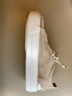
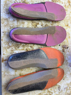
1/4 inch varus wedges applied to several OTC insert

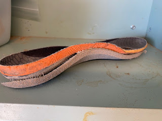
A closer look at the varus wedge applied along the medial aspect of the insert

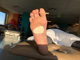
     Pronation is a normal function of the foot (with a similar motion described for the wrist). The foot pronates as you hit the ground creating good motion to absorb shock, and loosening the foot to adapt to the ground. Therefore, pronation is a normal and necessary function for the body.
     I just ran a workshop to 6 students at the California School of Podiatric Medicine at Samuel Merritt University. Two of these students were what you call excessive pronation where the motion of pronation lead to significant arch collapse and knee instability. One of these had the instability limited to the lower extremities. But, the other student also had trouble with his hips and back where the foot pronation destabilized the entire body. 
     Therefore, if the pronation motion is too much, too prolonged in gait, or too rapid to decelerate, symptoms can occur from the foot up. Since there are over 20 symptoms tied to the pronation syndrome, patients tend to pick on their individual weak spots. One excessive pronator can present with posterior tibial tendonitis, another plantar fasciitis, another knee or hip pain, up to the spine and upper extremities. 
     What does this have to do with varus wedges? A 1/4 inch varus wedge makes a typical 4 degree change in the pronation for a patient. The varus wedge can be just a heel wedge, but will not help as much for a runner when they go up on the ball of their feet. 
     Athletes in general are in my office complaining about their knee, ankle or foot. But, I like to talk to them about other problems they have to either a lesser degree, or significant past problems. They may have had serious bouts of plantar fasciitis, shin splints, achilles, etc, but just not now. 
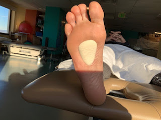
     One of my patients presented with posterior tibial problems and was very pronated. OTC arch supports and one pair of custom orthotic devices in the past were either painful to wear or did not fit into her shoes well. We spent some time over the next year designing comfortable but stabilizing orthotic devices. Over the next year I remember her counting over 10 areas in her body, mainly lower extremity, that did not hurt anymore (they were all part of this pronation syndrome). 
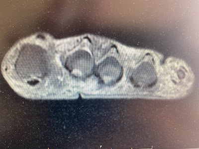
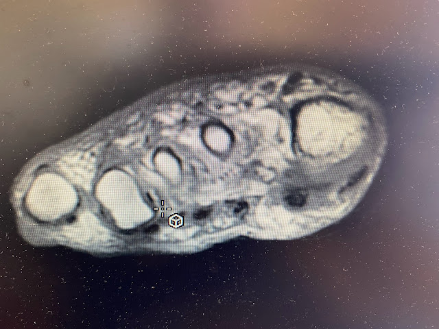
     Therefore, if you have some symptoms, or if your patients have symptoms, that you think could be tied to this syndrome of over pronation, applied a varus wedge as small as a varus wedge to one pair of shoes and see if the symptoms improve. It could be as vital as an MRI. 
     In the next few days, I will do a video to show how you can use an old shoe insert, cut the medial one inch off, apply it to another full insert to create a varus wedge. I sure hope this helps. Rich 

Friday, March 11, 2022

Beware of Subtle Xray Signs in the Foot


This image is of the 2nd toe (right) and 3rd toe (left).

     If you look at the major joint demonstrated (called the proximal interphalangeal joint), you can tell that there is a subtle cloudiness of the 3rd toe joint that is not in the 2nd toe joint. Since it was the 3rd toe that was the most sore and swollen from a run in with a furniture leg at midnight, and since the xray report was negative, I decided to get a lateral view of the toe involved. This is technically difficult as you have to get all the other toes out of the way so that there is no overlap. 


Here the lateral 3rd toe x-ray demonstrated a chip fracture off the base of the middle phalanx of that 3rd toe. I still remember our xray professor, Dr David Coulter, pointing out these subtleties, and I thank him at least once a week. 

Thursday, March 10, 2022

Hallux Rigidus: Surgery, No Surgery, or In Between

Here is the right big toe joint of someone whom has some minor DJD
(Degenerative Joint Disease AKA Wear and Tear)

Here is the left big toe joint of the same person with significant DJD

     Hallux Rigidus for many is a painful arthritic big toe joint. Patients can present with pain for many years or recent. Typically, like any sore joint, you can use common sense and get the joint comfortable. You may be just holding off the inevitable, the surgical knife, but who says that this is not worthwhile. 
Surgery is not without its problems. Most surgeries last 10-15 years and then have to be redone. If you fuse the big toe joint, you may not have big toe joint pain again, but you have totally messed up the normal pattern of movement. Our body must compensate for limping in pain, and it must compensate when a major joint is locked up permanently. 
     There are so many thoughts that run through my mind with this patient. One concerns why is the left side more broken down. We could discuss this for hours. Commonly, the left foot in our predominately right handed society gets beat up more. It is our support foot or support side that always takes more load in some way or another. Yet, podiatrists love to look for the nuisances to a pain syndrome like this. What also may put more pressure on the left big toe joint? Asymmetrical pronation is one, where the pronation or arch collapse places incredible stress on the big toe joint. Tight achilles and hamstrings, long leg syndrome, bone structural differences between the two sides, etc, all can place more force on the big toe joint which slowly and gradually collapses under this pressure. 
     Having been in a sports medicine and biomechanics practice my whole life, I have come to appreciate a non-rush attitude into surgery. Get more than one surgical opinion, and do not tell one surgeon what the other said. Find out if anyone can treat your problem conservatively. It is great to find out if there are mechanics that aggravate the stress at the injury that can be reversed. It is also great just to calm down the inflammation and relax any irritable nerves. 
     The last point today: treat the patient, not the xray. 


Sunday, January 30, 2022

Adding Stiffness to the front of a Shoe: Help for Many Problems in the Front of Our Feet


     My retired partner, Dr Jane Denton, known world wide of the Denton Modification for over supination, continuously used this metatarsal stiffener. It is a 1/16 to 1/8 inch thickness of out sole material. All chronic pain in the area may be helped with this shoe modification. When patients tell you that they feel better as the shoe stiffness increases, this may be something that a shoe cobbler can add to a more flexible shoe. It should be done on both sides (even if the pain is on one side). It can always be removed if not helpful, or after the need for stiffness passes. 

1/8 inch Stiff Out Sole Material Applied to Add Stiffness for Painful Metatarsals


The Stiffness has to be created with 1/16 to 1/8 inch Material only. This increases someone's falling odds due to separating the ground from the foot, so has to be broken in to gradually. The patients that it works for are really pleased that they can wear some normal looking shoes. 

Monday, January 24, 2022

Chemotherapy Can Destroy Nails and Hair: Ice to the Rescue

https://myhealth.alberta.ca/health/AfterCareInformation/pages/conditions.aspx?hwid=acj1006

Many of my patients have had to have chemotherapy and their nails have never been the same. Using an ice bath, head cap, etc, while having the chemotherapy infusion, can greatly help some patients. The theory is that you freeze the hair or nail cells while the chemo is working, thus preventing damage. Thank you Susan for bring this up to me. 


Squeaking Orthotics: JSuede helps

My small lab at Saint Francis Memorial Hospital has a roll of JSuede, from JMS Plastics, for the patients complaining of squeaking from the orthotic rubbing against the synthetics of the shoes


Here, both the bottom of the orthoses and the top of the JSuede is glued and stuck together.
This very thin material can help without increasing bulk too much

Sunday, January 23, 2022

Virtual Walks are really fun!!!

Totals after the first 5 days (out of 9) in this Virtual Walk

     The last 9 days I have been virtually walking from Canterbury England to Rome, Italy. 34 people from mainly England are participating. We made it the 1200 miles to Rome and are starting to head back. All of our miles count towards the total walked, but they also tell us our ranking among the other walkers. I have moved from 3rd to 6th place over the week. I am a competitor by nature, and this has been fun and motivating to walk daily more than 10,000 steps over 5 miles. I have one more walk today and it is over. We will not make it back to Canterbury as a group, but I will not have to fly from anywhere either. 
     The website I am using for this virtual walk is UltraBritain.com. It is a charity event to raise money for the maintenance of this historical walk. Canterbury to Rome has been a well travelled route since 500 AD. It goes by boat across the English Channel and then through France and the lowest part of the Swiss Alps. 

Friday, January 21, 2022

Adding Stiffness to the Front of an Orthotic Device

https://jmsplastics.com/product/j-suede/


     The image above was taken after I applied a very thin piece of suede leather. This is stretched before tacking down to stiffen the bend across the metatarsal heads. This is a very useful technique when trying to treat metatarsalgia symptoms, Morton's neuroma symptoms, hallux limitus and rigidus symptoms, and all without adding alot of bulk in that painful area. 


Wednesday, January 19, 2022

Good Rx: A Good Way to Get Some Prescriptions at a fraction of the cost


https://www.goodrx.com/how-goodrx-works

     This company has saved hundreds of my patients a lot of money while buying prescriptions. Please be aware of it. 

Monday, January 17, 2022

Sunday Night Musings of a Personal Nature

A View of Sunset in San Francisco from my walk tonight!

     I am working on a book for the last five years. Months will pass and not a word is placed on a page, and then I get really productive. I am writing this podiatry book for my students at the California School of Podiatric Medicine in Oakland, California. The Podiatry school is part of the incredible medical graduate programs run through Samuel Merritt University. 
     The book title is Practical Biomechanics for the Podiatrist and really focus' equally on both basic treatments and more sophisticated. In medical school, the emphasis is on teaching high tech, so I plan on cornering the market with KISS techniques (Keep It Simple Stupid). 
      I am basically trying to teach the thought process of helping patients throughout the whole injury. My book, however, has gotten too big, and too time consuming, that I have now broken it down to 4 books with book 1 coming out this year. Wish me luck!
     RETIREMENT!! Well, I was going to retire at 65, and now I just turned 68. So, from today on, I am sticking to my #70 birthday on 1/9/24. At my age, you worry about illness' that will suddenly bring you down at any age. I started working at Saint Francis Memorial Hospital when I was 27 years old!! Long time in one place, but I have loved it. 
     Unfortunately, Saint Francis is not finding a replacement for me. Most of my present patients will have to find someone over the next 2 years. It will be a great 2 years!!


Sunday, January 16, 2022

Lecturing: A Way of Giving Back

     Of course, I am the handsome guy to the left as you look at the photo!!LOL
Why am I showing you this? I am at the end of my career, and giving back as much as I can is very very important to me. This photo is for some online presentations in Spain. I will talk to almost anyone, in fact any one, who will listen. I believe in my careful blend of biomechanics and sports medicine. Patients get better, or get a better understanding of their problem. Patients and I are partners in healing. 
     This year I am lecturing at the California School of Podiatric Medicine through April, and then again in October and November. I am also lecturing at our state's meeting in Los Angeles in June. I may give an online lecture in Oman in March or April, and I hope to be invited back to New York next year. 
     What sticks in my side always was that one of my mentors, Dr John Weed, died at 54 while working at a busy office. I watched him work for over 13 years, a truly gifted and loving individual. He was excited about retiring some day so he could write of all his discoveries. Podiatry truly missed out. He died 30 years ago, and I still miss him a lot. As I try to give back to my profession, I wish he had been given the opportunity. 


Thursday, January 13, 2022

Common Foot Pads used to Off Weight Sore Areas


Adhesive Felt used to off weight ball of foot
Typically the padding is placed on the undersurface of the shoe insert

Adhesive Felt used to off weight the 2nd and 3rd metatarsals where they bear weight


Wednesday, January 12, 2022

MRIs: Their Usefulness in Patient Care of the Foot


     Those of you whom regularly read my blog know that I love MRIs and even serial MRIs when I am following an injury. Unfortunately, the quality of the MRIs is getting poorer, and the amount of the foot that is imaged is becoming less and less. That does not mean you can not get great MRIs but you may have to ask for the highest Tesla (magnetic strength) in your area. 
     Why is a negative MRI just as important in general as a positive MRI? Negative MRIs rule out bone injuries, ligament injuries, and cartilage injuries. Negative MRIs suggest that a patient may have nerve pain if all other causes of the problem have been ruled out. 
     In the MRI above, even though you are given only one of the 144 slices, the 2nd metatarsal phalangeal joint had a lot of inflammation. That is the white you see under the 2nd and 3rd metatarsal heads. Other slices confirmed no plantar plate injury (the ligaments under the metatarsal heads at the ball of the foot area). 
      When you see inflammation, you have to think that this area is being stressed. The inflammation is the bodies' way of healing itself. Inflammation brings in nutrients for healing, therefore very important for healing. Inflammation though is always an all or nothing response. The body feels like it is under attack, and it will send in the calvary, armed forces, and Knights Templar (even Luke Skywalker). All or nothing!! Fight or flight phenomenon. The guist is that the inflammation can be more than it needs. Chronic inflammation, still sitting around from a relatively minor injury, still hurts alot. The patient then wonders why they have not healed, even when they have. I see this phenomenon over and over.
     Inflammation also means something may be happening internally, like micro-tearing in the ligaments. I can not see it in the MRI, but the patient can not get rid of the inflammation by any means (PT, contrast bathing, ice massage, acupuncture). 
     In this individual, negative MRI except some inflammation (radiologists call this edema). Initial treatment to address the inflammation alone was nonproductive. Treatment switched to restricting joint motion with Budin Splints, and the pain got better real quickly from there. 
     I tell my patients that positive or negative MRIs are data points in their treatment plan. We have to react to the information, make changes when they are not improving, and follow the progress month by month. 
     

Sunday, January 9, 2022

Happy Birthday Me!!

     Yes, it is my birthday today. If I was born yesterday, I could of shared Elvis Presley's big day. But no, I got Richard Nixon. It was a cool birthday until the world found out he was a crook (along with other things). But, I digress!
     Thank you all for reading this blog. I can not believe the blog is also celebrating its birthday in March. 13 years for the blog, and 68 for me!! 


Saturday, January 8, 2022

Customizing an OTC Insert: The list is endless and limited by our Imaginations!!

Sole is one of the common OTC arch supports I recommend in my office

In this case, the front was just too hard for the patient so I removed it for softer material

The cut or transition was just behind the metatarsal heads (ball of the foot)

Here the Arch only part with be glued onto a full leather top cover. 

Using the original front, the leather can be trimmed to be an exact replica of the original


Now a soft material like 1/8 inch poron is applied as the new front


Thursday, January 6, 2022

Bunion Protection in Tight Shoes

Here is 1/4 inch adhesive felt (which can be used for 3-4 days) is placed behind the bunion prominence (never over) while wearing shoes

Wednesday, January 5, 2022

Metatarsal Pads: Proper Placement

Hapad Longitudinal Small Metatarsal Arch Pads are my preferred Pad.
These pads spread the weight over a large area and can be thinned by peeling layers off.
Hapad Metatarsal Pads can be used in 2 directions just before the sore area on the foot. This is the common direction.

This is a common modification of the small Hapad metatarsal pads with the thickest part under the 2nd metatarsal head. These adhesive felt pads also can be easily adjusted.


Sunday, January 2, 2022

Soccer Cleat and Foot Pain


     One of the common problems I see when treating soccer players is that the painful area is right over one of the cleat positions. I need a convincing argument to explain that they need to change their cleats or shave down at least half of the offending cleat. The fear of losing traction or gaining instability by removing at least part of the cleat is often the reason they would prefer to switch the type of cleats that they wear in the long run. 

Saturday, January 1, 2022

Plantar Plate Injuries Algorithm


How Are We Built to Function? Loose and Unconnected, Perfect, or A-little of Both


     Daily in my evaluation of the structure and function of patients that present to my office, the patients can feel something is off. It can feel a little like these Tinker Toys to both patients and new doctors learning the trade. There are so many rules of what is ideal, but patients are typically not seeking to be ideal. They only want their symptoms to feel better, and their bodies to feel more stable or in better alignment. 
     I am a huge proponent of gait evaluation. During gait evaluation, many of the uniqueness' of an individual can become obvious. As you watch someone walk or run, you will see patterns of function that may be related to symptoms or may be related to great health. When patients come in with a problem, and I watch them walk and/or run, I think they are a bit disappointed when I tell them that they are fine (stable, smooth, efficient). They are looking for answers to their issues, but it can not be found in gait. Other patients come into the office with foot pain, and when I watch them walk, you can tell why their knee or back hurts and how you can help. They had not even mentioned these areas because they were in a Podiatrist's office. 
     When I teach podiatry students, I always emphasize to walk every patient walk or run as the clues can be literally amazing. 

Monday, December 27, 2021

Very Tight Tendons: Consider Heat Ice Prolonged Stretching


This patient has a very tight achilles tendon. The prolonged heat ice routine is based on a study done at Temple University years ago. The goal is to 3 times per week hold a stretch throughout a 30 or even 45 minute period. Heat is utilized here in the form of a heat pack for 30 minutes and this is followed by an ice pack for the last 15 minutes. Therefore, it is a 2:1 ratio of heat to ice, so 20 minutes of heat followed by 10 minutes of ice, or 15 minutes of heat and 7.5 minutes of ice. You can see it is a two man job! I get great results within a few weeks (probably near the 10th time landmark). The days the patient is not doing this they still need to stretch 3 times normally. 


Sunday, December 26, 2021

Nerve Pain: Where Does It Come From?


     This image is of a T1 MRI section across the front of the right foot. Above my sensor marker, there is an obvious Morton's Neuroma that may be the complete cause of this patient's pain. Remember, we need to always ascertain if the pain is mechanical, inflammatory, neuropathic (like Morton's Neuroma), or a combination of these 3 factors. 
     In this foot, the section shows a typical low lying 4th metatarsal head with a very thin fat pad. This allows these plantar nerves on the bottom of the feet to get beat up too easily. The Morton's Neuroma can develop from this constant abuse over years. Why they begin to become symptomatic, when they are fairly large, is anyone's guess? 
     The onset of MRI technology now 35 years ago did teach us an incredible thing: Not all Morton's Neuromas hurt as they were found in patients where they never had nerve symptoms. Therefore, it is important, even in the face of an obvious MRI documented symptomatic Morton's Neuroma, that we make sure that the pain is completely driven by this enlarged nerve.
     The 3 sources of pain: mechanical, inflammatory, and neuropathic, also come the 3 avenues you can treatment patients symptoms: mechanically, anti-inflammatory, and nerve desensitization. So, we begin treatment with mechanical off-weight bearing pads, icing and contrast bathing, and neural flossing or acupuncture, etc. And, we follow these simple treatments with others based on the patient response and subjective feeling on what is helping. 
     Morton's Neuromas, as well as other nerve conditions like Tarsal Tunnel, have the added caveat that the majority of symptoms do not originate in the foot. This implies, and is very true, that treatment alone of foot nerve pain at the foot may not be successful. You typically should include in any Morton's Neuroma workup and treatment getting consults on the low back and spine in general. Think about the concept of Double Crush, where the nerve is only painful when irritated at least in two places, with no symptoms if you remove one of the two areas of irritation. 
     

Sunday, December 19, 2021

Podiatric Question #1

Practical Biomechanics Question #1: What position is the ankle in when it is the most relaxed, or the least stressed?


The ankle is the most relaxed at a 20-30 degrees plantarflexed pointed position and in Subtalar Neutral (neither pronated or supinated).

Plantar Fascia Healing of Partial Tear: 3 MRIs (6 months apart)

Sept 2020 Medial Slip Plantar Fascia Partially Torn

April 2021 Some Darkening (Consolidation Beginning) of Plantar Fascia But Tear Remains

December 2021 Good Consolidation Noted


Big Toe Pain: Email Advice

Hi Dr. Blake,

     I found your blog after dealing with chronic toe pain for the past 4 years. It started in 2017 when I was trying to stay healthy and did a bunch of yoga. I ended up doing a lot of hand stands and every time I would end I would plant my right great toe to the ground. Didn't start bothering me until I did some minimalist incline hiking. 
     Limped for a few weeks and had some serious pain below my great toe, gradual felt better, never 100%. I wound up on a beach a month or so later and have never been the same. I walked a little and then all of a sudden couldn't plant with my great toe due to the pain. limped for weeks and finally saw a specialist. 
     I was initially diagnosed with hallux rigidus, seemed kinda crazy. Luckily I found hokas and was able to walk normal and have been in them ever since. Saw multiple other doctors and had an MRI a year later. 
     I had a sesamoidectomy after the surgeon thought there was an unhealed fx that may have been causing the issue. The certainly didn't help. Have seen multiple surgeons since and just had a 3rd MRI, this time it showed a possible partial tear through the plantar plate. 
     I initially thought this was the injury but was told it wasn't because there was no instability and the MRI read didn't mention one. But now since it did, I have a feeling that was the issue all along. 
     Anyways I am reaching out for advice. Would a partial plantar plate tear heal after 4 years? Dr. Blake's comment: Probably not, but when you had the sesamoidectomy didn't they see a tear, or make any comment? Odd. You probably have to at least send me the MRI reports: all 3 to look at. 

What options are out there for me? Dr Blake's comment: The treatments are many to control pain and allow activity including: Hokas, spica taping, off weight bearing orthotics, avoiding activities that cause pain over 2, etc. However, the diagnosis is what is crucial and 3 MRIs should be fairly conclusive. 

 I have been wearing hokas for the past 4 years and have not participated in any physical activities that I like or even ran since 2017. Really hoping to change that. Any recommendations or advice would be greatly appreciated. Thanks - 

Monday, December 13, 2021

Posterior Tendon Dysfunction: Email Advice

Hi Dr Blake,

I’ve been reading your blog all day, thank you for proving this invaluable resource. I’ve been doing lots of research into posterior tibial tendon dysfunction and your site is a wealth of knowledge.

So I’ve been having some PTTD type problems. I ran an 73 mile ultra in July this year and really damaged the posterior tibial tendon. So much so I haven't been able to run properly since the injury. I’ve done some downhill walk running which was ok 5km ish.  I have undergone reasonably intense physio protocol since September and I am now at the point where I am pretty much pain free day to day. I walk to work 45 minutes each way without pain, or with mild 1 or 2 pain on pain score.

Unfortunately my medial ankle started making a snapping sensation, which is worrying me. It feels like a ligament, possibly the PTT is snapping across the medial malleolus. Although it could be something to do with the deltoid ligament, this is where I notice the snapping sensation most, I am not sure of the cause.  My PT gave me stretches and strengthening exercises, but the snapping only started occurring after I started rehabbing the injury. Now I am at the point where I am not sure if the tendon is loose and that’s causing the snapping or if it’s tight and that’s causing the snapping. There’s never an audible click as much, it’s more of a sensation you/I can feel. It can only be felt when the foot is dorsiflexed and inverted from the normal position upwards and inwards in that order. I sometimes notice the snapping when walking up hill. If the foot isn’t dorsiflexed and inverted then the snapping doesn’t happen.

My PT has helped, but isn’t a specialist in foot injuries, I can’t see a podiatrist until February and I am a mountaineer keen to get out as soon as possible. I was wondering if you had any advice regarding the snapping or ways to make the snapping stop? Everything I’ve found online in relation to snapping talks about the peroneal tendon, but that’s on the outside of the foot, and isn’t the source of my problem. I can’t find anything that specifically helps my inner foot snapping issue.

Best regards,

Dr Blake's Response:
     Thank you so much for your question. This occurs to my patients all the time, so I am hopeful you can resolve this. There are alot of things that happen when the ankle collapses inward in the case of posterior tibial problems. Tendons get stretched, ligaments stretched, and inflammatory builds up in the the joints and soft tissues. Typically, if you are non weight bearing, and you move your ankles around in all directions, if a tendon or ligament was loose, you should be able to duplicate the snapping sound, and see something dramatic happen. In your case, it sounds like it is only with weight bearing, where the snapping is due to fluid trapped in joints and ligaments. The snapping sound is the fluid escaping as you move much like the snapping of finger joints. I would attack this hard with anti-inflammation, even if you can not see the inflammation. Icing twice daily, contrast bathing each night, 3 advils three times a day, etc. Then get back to me in one month and remind me of this blog post (attach the link). Rich 

Monday, November 29, 2021

Treating Nerve Pain at the Foot: Algorithm


     I treat many patients with nerve pain, some as the primary injury like Morton's Neuroma, and some as secondary nerve hypersensitivity develops following an injury. This algorithm can help the patient with nerve pain review their treatment and discuss other options here with their doctor. 

Sunday, November 28, 2021

Plantar Fasciitis: Treatment Algorithm


     The treatments are typically non-painful stretching as outlined in this video. 


Weight transfer concerned getting the weight into the arch with an orthotic device. This is successful when most of the pain is in the heel or ball of the foot areas. 


Podiatrists always love to recommend the Rolling Ice Massage technique 5 minutes twice a day to get the inflammation under control. 


Tuesday, November 23, 2021

The 1 Day and 2 Day Pain Level Increase Rules of Rehabilitation

     When I am rehabilitating a patient, we always talk about good and bad pain. It is so important for the patient to know the difference. And, it is very important for me to know where their pain is in my rehabilitative process. 


     When you are rehabilitating a sore area, and you are progressing the patient through the 3 Phases of Rehabilitation, increased stress to the area is applied routinely every other day in activity progressions. This is typically Monday, Wednesday, Friday or Tuesday, Thursday, Saturday. You have kept the pain between 0-2 successfully, but this increased stress (important for healing and return to activities) will elevate the pain for a period of time following. The goal during rehabilitation to keep the increased tissue soreness within 2 days or the 2 Day Pain Level Increase Rule. Therefore, if I am starting a more strenuous activity, like progressing to the Walk Run Program, or allowing small jumps in ballet, the increased pain needs to be contained within 2 days and not linger more. If you exercise on Monday, by Wednesday's start you should be back to square one even if Tuesday showed increased pain. 
     The One Day Pain Level Increase Rule is for the maintenance program. You are back at your normal level. You are exercising at a good level 3-4 times per week. Any increased pain after an activity is normal as long it is not during activity, not increasing in severity week to week, and contained with one day. 
   It takes alot of understanding to excellently rehab a patient, or yourself, of these 2 rules to avoid serious setbacks. Good luck 

Saturday, November 6, 2021

Big Toe Area Pain: Years after Healing Sesamoid Injury

Hi Richard,

I wanted to write to you because of pain in my big toe that has been bothering me for the past few weeks (about 5 weeks). Unfortunately, this pain is fickle and elusive, and very hard to describe.

My history:
- turf toe and sesamoid AVN in 2019-2020, which I recovered from thanks to your wonderful blog
- I was walking perfectly fine for a ~6 months
- played tennis one day and had pain the next day in my big toe, but a different one than I'd had before - it hasn't gone away unfortunately

The pain:
The pain is sometimes very sharp when I put weight on my foot right after getting up, but then immediately subsides. At first it felt like "pins and needles", but now it hurts more "traditionally". I can't tell where exactly my toe is hurting, but it definitely hurts when I press in the webbing between the first and second toe. My joint clicks a bit (though when it does, it is painless), and to be honest I can't remember whether it did that before feeling the pain or not. Moving my big toe doesn't hurt, I have full range of motion, my sesamoids seem fine. It occasionally hurts when I compress my first phalanx with my hand, I think it's called the "proximal phalanx of the big toe". Overall the pain tends to be better in the morning, and worse after a whole day of walking.

Please let me know if you have any leads, because I admit I'm quite perplexed by my current condition!

May God preserve you and your family in this difficult time. 

With all my consideration


Dr Blake’s Comment: Thanks for your email and glad the sesamoid AVN finally healed and you were back to good activity. Some of the symptoms are definitely nerve, like the pins and needles and pain that comes on and disappears quickly. In your case, it could have just be the body trying to tell you that something is amiss that needs protection. Your original injury was to the big toe joint. Previous joint injuries tend to pop up from time to time. Like you are doing now, you have to take them seriously, but they represent that the joint is not perfect. Imperfect joints (for me they are the left ankle, right knee, low back, and right shoulder) from old injuries are generally alittle stiffer (so they can get jerked easier) and the body’s reaction is quicker (from nerve memory). 

     So, what does this all mean? Typically, we are not dealing with anything serious if the range of motion is normal and there is no swelling, black and blue, or redness. Also, it is a great sign that there are times of the day that it does not hurt at all. These aggravations of old injury areas need to be treated seriously since this joint is a weak spot for you. The 3 areas to address are mechanics, probably spica taping or bunion toe separators to start, inflammation (so icing and contrast bathing once or twice daily), and nerve hyper-sensitivity (neural flossing with non painful joint motions, and non painful massage for 2 minutes twice daily. This should be done for 2 weeks, and then based on the response, either lessen your treatments or increase them in some way. 

     I hope this makes sense. Rich