Total Pageviews

Translate

Followers

Saturday, March 12, 2022

Pronation Syndrome: The use of varus wedges for treatment and diagnosis

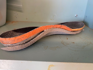
1/4 inch varus wedges applied to several OTC insert

A closer look at the varus wedge applied along the medial aspect of the insert

     Pronation is a normal function of the foot (with a similar motion described for the wrist). The foot pronates as you hit the ground creating good motion to absorb shock, and loosening the foot to adapt to the ground. Therefore, pronation is a normal and necessary function for the body.
     I just ran a workshop to 6 students at the California School of Podiatric Medicine at Samuel Merritt University. Two of these students were what you call excessive pronation where the motion of pronation lead to significant arch collapse and knee instability. One of these had the instability limited to the lower extremities. But, the other student also had trouble with his hips and back where the foot pronation destabilized the entire body. 
     Therefore, if the pronation motion is too much, too prolonged in gait, or too rapid to decelerate, symptoms can occur from the foot up. Since there are over 20 symptoms tied to the pronation syndrome, patients tend to pick on their individual weak spots. One excessive pronator can present with posterior tibial tendonitis, another plantar fasciitis, another knee or hip pain, up to the spine and upper extremities. 
     What does this have to do with varus wedges? A 1/4 inch varus wedge makes a typical 4 degree change in the pronation for a patient. The varus wedge can be just a heel wedge, but will not help as much for a runner when they go up on the ball of their feet. 
     Athletes in general are in my office complaining about their knee, ankle or foot. But, I like to talk to them about other problems they have to either a lesser degree, or significant past problems. They may have had serious bouts of plantar fasciitis, shin splints, achilles, etc, but just not now. 
     One of my patients presented with posterior tibial problems and was very pronated. OTC arch supports and one pair of custom orthotic devices in the past were either painful to wear or did not fit into her shoes well. We spent some time over the next year designing comfortable but stabilizing orthotic devices. Over the next year I remember her counting over 10 areas in her body, mainly lower extremity, that did not hurt anymore (they were all part of this pronation syndrome). 
     Therefore, if you have some symptoms, or if your patients have symptoms, that you think could be tied to this syndrome of over pronation, applied a varus wedge as small as a varus wedge to one pair of shoes and see if the symptoms improve. It could be as vital as an MRI. 
     In the next few days, I will do a video to show how you can use an old shoe insert, cut the medial one inch off, apply it to another full insert to create a varus wedge. I sure hope this helps. Rich 

Friday, March 11, 2022

Beware of Subtle Xray Signs in the Foot


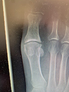
This image is of the 2nd toe (right) and 3rd toe (left).

     If you look at the major joint demonstrated (called the proximal interphalangeal joint), you can tell that there is a subtle cloudiness of the 3rd toe joint that is not in the 2nd toe joint. Since it was the 3rd toe that was the most sore and swollen from a run in with a furniture leg at midnight, and since the xray report was negative, I decided to get a lateral view of the toe involved. This is technically difficult as you have to get all the other toes out of the way so that there is no overlap. 


Here the lateral 3rd toe x-ray demonstrated a chip fracture off the base of the middle phalanx of that 3rd toe. I still remember our xray professor, Dr David Coulter, pointing out these subtleties, and I thank him at least once a week. 

Thursday, March 10, 2022

Hallux Rigidus: Surgery, No Surgery, or In Between

Here is the right big toe joint of someone whom has some minor DJD
(Degenerative Joint Disease AKA Wear and Tear)

Here is the left big toe joint of the same person with significant DJD

     Hallux Rigidus for many is a painful arthritic big toe joint. Patients can present with pain for many years or recent. Typically, like any sore joint, you can use common sense and get the joint comfortable. You may be just holding off the inevitable, the surgical knife, but who says that this is not worthwhile. 
Surgery is not without its problems. Most surgeries last 10-15 years and then have to be redone. If you fuse the big toe joint, you may not have big toe joint pain again, but you have totally messed up the normal pattern of movement. Our body must compensate for limping in pain, and it must compensate when a major joint is locked up permanently. 
     There are so many thoughts that run through my mind with this patient. One concerns why is the left side more broken down. We could discuss this for hours. Commonly, the left foot in our predominately right handed society gets beat up more. It is our support foot or support side that always takes more load in some way or another. Yet, podiatrists love to look for the nuisances to a pain syndrome like this. What also may put more pressure on the left big toe joint? Asymmetrical pronation is one, where the pronation or arch collapse places incredible stress on the big toe joint. Tight achilles and hamstrings, long leg syndrome, bone structural differences between the two sides, etc, all can place more force on the big toe joint which slowly and gradually collapses under this pressure. 
     Having been in a sports medicine and biomechanics practice my whole life, I have come to appreciate a non-rush attitude into surgery. Get more than one surgical opinion, and do not tell one surgeon what the other said. Find out if anyone can treat your problem conservatively. It is great to find out if there are mechanics that aggravate the stress at the injury that can be reversed. It is also great just to calm down the inflammation and relax any irritable nerves. 
     The last point today: treat the patient, not the xray.