Total Pageviews

Translate

Followers

Wednesday, November 30, 2022

Arch Height In Custom Orthotic Devices

The "H" is the High Point for the Medial Arch in a Custom Device

     The power of custom orthotic devices are so minimized by our society, and sometimes even by Podiatrists. The foot is the foundation of our bodies. If you have foot pain, you know what that means exactly when you can not walk. When you have ankle, knee, hip and back pain that has been improved by orthotic devices, you understand the importance of this foundation. 
     I just attended the 10th Annual Richard O Schuster DPM Memorial Biomechanics Seminar. We talked biomechanics for 2 straight days. For me, it was Podiatry Heaven. Those like me felt the same. Many of the speakers come from different approaches to Biomechanics which is all the better. You want your ideas and concepts to grow. 

Tuesday, November 29, 2022

Sesamoid Injury: Cortisone Shot or Not?

Hi Doctor Blake,

I stumbled upon your blog after lots of sesamoid research! And now I am asking for your help with my recovery... 

Long story short I’ve been off from dancing (just wearing special sneakers with dancer’s pads) for 7 weeks now for a micro fracture of my sesamoid. I just received an MRI with these results below (apologies if the translation from another language is weird):

Presence of bone edema of the entire medial sesamoid, with low T1 hypersignal T2 signal.  No fracture-separation of the bone, no sign of necrosis, and in particular no deformation of the bone surface.  Reactive joint effusion of the MTP 1. Light hallux valgus (confirm on radiography under load).  No sign of sesamoido-metatarsal osteoarthritis.  A little edema of hyperfunction of the plantar tissue in contact with the medial sesamoide.  No anomaly of the stabilizer of the sesamoid.  No abnormality of the hallux tendons.  Moreover no anomaly of other MTP and inter-capito-metatarsal spaces.  CONCLUSION: Appearance confirming medial sesamoiditis, without fracture or underlying necrosis.  Light hallux valgus (confirm on x-ray in charge)

I am seeing two different specialists in my country but now I have run into the problem of opposing medical solutions and I am not sure which to go with. 

One doctor recommends a cortisone injection and the other recommends oral anti-inflammatory medication. The doctor who recommended the oral medication told me that the cortisone injection is very dangerous for a sesamoid as it damages the foot’s natural padding, therefore exposing the sesamoid to further damage... 

The doctor who recommended the injection told me that the oral medication will not do anything to help the sesamoid and that all stories about sesamoid’s being made worse by injections are mainly myths and cannot be proven.

I am emailing to ask your advice on this debate? I am hesitant to get the injection because of all the mixed reviews online but it is a more “immediate” and localized option which is tempting. Do you have any feedback or success stories of oral medication? Or any thoughts on cortisone’s long term risks?

Any feedback you could offer would be greatly appreciated! 

My apologies for such a dense email, but your knowledge would be so helpful for me!

I thank you in advance for taking the time to read this email and I hope to hear from you soon.

Sincerely,

Here on this MRI image healthy bone is dark. The light tibial sesamoid indicates a healing response from the body. Hard to tell stress fracture (can not see) from bone bruise in these cases


Dr. Blake's comment: 

 I must side with the oral medications, which can not be taken before you dance (only after). Your MRI shows that the sesamoid has been slightly injured. Cortisone will mask pain for up to 3 days (short acting cortisone and not dangerous) to 9 months (long acting cortisone). Your dance career can not risk masking pain where the sesamoid injury worsens. You can ice twice daily and do contrast bathing every evening. You must float the sesamoid with Dr. Jill's dancer's pads of either one eighth or one fourth inch (3 to 6 millimeters). These can be bought on line and worn even walking around barefoot to protect the sesamoid. I have my patients get 2 lefts (both sizes) and 2 rights (both sizes) since the adhesive is on one side and at times you are wearing it on your foot, and at times you are putting it over or under inserts in shoes. Whatever you put on your injured foot, should be on the other foot for balance. I hope this helps. Rich 

Video On Designing Dancer's Padding for Big Toe Joint Problems

Sunday, November 27, 2022

Video from Dr Richard Blake on Injury Rehabilitation General Rules

https://prezi.com/v/view/wyPiHlcIQ9LKeHoi88I2/

POSE Running: Video on the Basic Components

https://youtu.be/shwi2MfxSok

     I have treated runners of over 40 years. The POSE running concept is a sound one. I would just at a lecture by Dr Jim Losito in Miami. Dr Losito is an expert in sports medicine and past president of the American Academy of Podiatric Sports Medicine. He is a huge proponent of POSE running for injury prevention and running efficiency. This blog and accompanied video is merely to say "yes" to have you learn the basic concepts in other videos. 

Friday, November 25, 2022

Love Your Podiatrist: Consider Book 1 or Book 2 for Practical Biomechanics for the Podiatrist as a Christmas Present


They will think you are very special!! Happy Holidays!! Rich 

Shock Absorption Problems: A Common Cause of Lower Extremity Problems

     Many of my patients have problems with shock absorption. This excerpt from Secrets To Keep Moving ebook mention a few. We watch patients walk and see if there are any problems, then we watch them run (if they run). We listen to their histories on how they were injured. I am looking for internal vulnerabilities (reasons that the foot or gait causes poor shock, or for external stress like poor shoes, excessive downhill walking, etc. As podiatrists we focus too much on over pronation, when poor shock absorption, limb length discrepancy, inherent instability, weak and tight muscles, over supination, and forefoot deformities can be equally the causal effect of an injury or pain syndrome. Rich 

 Poor Shock Absorption
  • Tibial Stress Fractures
  • Knee Pain

  • Calcaneal (heel) stress fractures
  • Metatarsal stress fractures
  • Heel Bursitis or Heel Bruise
  • Femoral Stress Fractures
  • Where there are many causes of poor shock absorption, patients who have excessive supination always have poor shock absorption. You need contact phase pronation to absorb the shock at heel strike.
 
Sometimes even my smallest slightest patients can walk very heavy and have poor shock absorption

Thursday, November 24, 2022

5th Metatarsal Fractures: A Special Breed (General Rules)

Fifth Metatarsal Fractures: A Special Breed (by Richard Blake, DPM)


    The top 10 initial treatments for 5th metatarsal fractures are:


  1. X ray evaluation to decide on surgery vs conservative care.
  2. If surgery, protocol to be set by surgeon and not the purpose of this writing.
  3. If conservative care chosen, some form of immobilization for 8-12 weeks is typically done based on injury (Immobilization Phase). You want to get the pain level between 0-2 with 2 weeks, and maintain that during the entire rehabilitation.
  4. During the Immobilization Phase, lower limb strengthening with some cardio should be orchestrated by a physical therapist. Even one legged stationary bike is very beneficial.
  5. Bone health is analyzed with dietary calcium and Vit D3, consideration of a bone density screen, and typically healthy diet.
  6. Transition period from cast to no cast, with or without surgery, can be very difficult. Custom orthotics with full lateral arch support very helpful. At times, extra big shoes during the transition can be purchased so added padding/accommodation can be used.
  7. When not using a permanent cast, 24/7 compression bandages, ice pack 15 minutes twice daily, contrast bathing each evening, as much as possible elevation, 3 times daily 3 minute self massage for desensitization and swelling reduction, and hourly pain free ankle circles are initiated.
  8. Weight bearing for bone mineralization, even in casts or boots, is done as early as safe (Good Pain vs Bad Pain)
  9. All fifth metatarsal fractures, except a few styloid process avulsion fractures, should have a Exogen Bone Stimulator for 6 months (when insurance allows).
  10. Follow up xrays need only be done when symptoms plateau or worsen. As long as the patient makes steady, gradual, progress, it is better to base improvement on function, not x ray or palpable tenderness.

    A. Fifth Metatarsal Fractures: Non Jones Type


These images are from a patient of mine that is almost 3 months post injury and her x-rays show a wide gap still. Here are all the thoughts that are meandering through my brain.


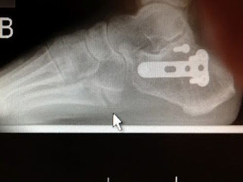
Here is the standard Lateral view with quite a large gap noted
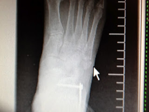


A Jones Fracture to the Fifth Metatarsal is normally 1 inch closer to the toes. This AP view still shows some displacement.
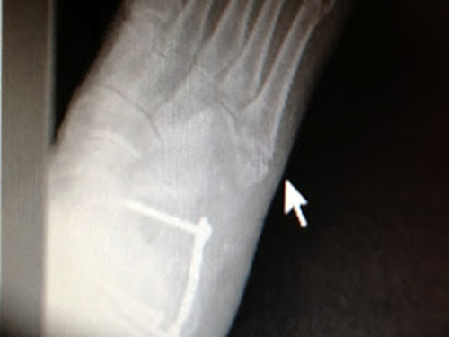


This Oblique view makes the fracture clearer and you can see if the fracture line goes into the joint of the 5th metatarsal/cuboid.


You 
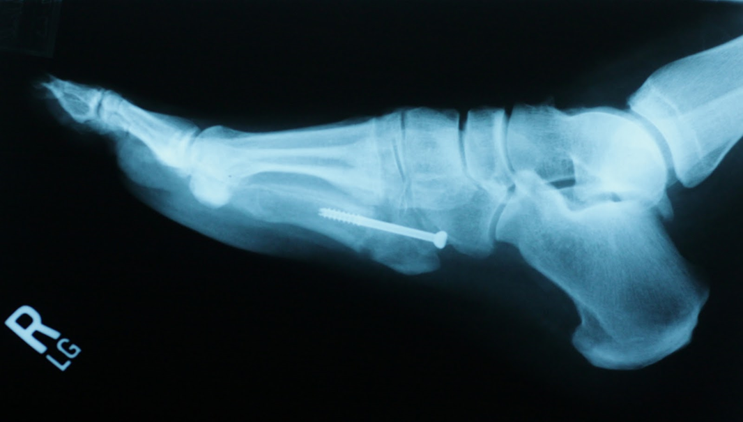
can see in this post Jones fracture repair xray that the Jones fracture is further forward than a 5th metatarsal avulsion fracture. 

This 5th metatarsal avulsion fractures following some inversion twist of the foot are typically under treated. Because they do not have the stigma of a true Jones fracture (historically more serious), they can be less aggressively treated. Sometimes this is okay, and sometimes not. Again, the goal is to create a pain free environment, which I believe has happened. X-rays for foot fractures, since the healing normally takes place internally first, cannot really reflect the strength of the bone. But, I do not like the gap and I do not like the fact that the joint is involved (possibly future arthritis).


    So, what are all the steps we need to make happen?


1) Establish a pain free environment if not already occurring

2) Make sure Bone Strength is good (questioning about Vit D3 and Calcium, bone density, healthy diet)

3) Stabilize the fifth metatarsal with orthotic devices, accommodative padding, and kinesiotaping (there are special techniques in orthotic devices for the outside of your foot)

4) Set workout goals that do not over stress this area

5) Avoid anti-inflammatories since they can slow down bone healing

6) Ice Pack 15 minutes twice daily, and contrast baths once daily to reduce inflammation

7) Due to the gap, seek approval for Exogen Bone stimulator

8) Have patient talk to a surgeon to find out what the process of fixing if the above does not work (this keeps the patient well informed)

9) Advise on possible future arthritis

10) Only get future X-rays if treatment has plateaued (there are many cases of pain free non healing) since current healing of the bone is not reflected well on x-rays.


Happy Thanksgiving from Drblakeshealingsole


     I hope this year brought you many things to be thankful for. Most of those things are normally nothing you buy. They are visits with family and friends. They are moments of elucidation when struggling with a concept. They can be the phone call from a friend to comfort you or a good book. They can be some music that unexpectedly wells something up in your heart.
     Many of my patients are struggling with physical and emotional issues. I am thankful I can be there for them. I am thankful for the silence in nature or when reading a book. I am so grateful when my favorite team wins a big game. And, I am thankful for you, my readers. I am humbled by the thought that my words can be of help to you. Have a wonderful day!!

Wednesday, November 23, 2022

Neuro One: Topical Medication for Peripheral Neuropathy

https://youtu.be/7odegu-00pk

Here Caroline Jordan helps pitch a very good topical nerve salve called Neuro One. Partially invented by Dr Arlene Hoffman, this cream like Neuro Eze, works in some 50% of my patients who complain of nerve sensations in the feet. It is of course part of an overall plan. 

Hallux Rigidus: Position Paper on Treatment

     Hallux Rigidus means that there is joint limitation of the first metatarsal phalangeal joint to a degree of 30 degrees dorsiflexion motion or less non-weightbearing or 10-15 degrees or less weightbearing standing or walking. Hallux Rigidus is normally associated with significant arthritic changes in the first metatarsal phalangeal joint. These joint arthritic changes can develop from old trauma or long standing microtrauma at the joint level eventually leading to observable joint changes. These joint changes can be spur formation, joint narrowing, and other signs of cartilage loss. When a patient presents with pain in the big toe joint, in the present of a joint arthritic condition, too often the first treatment is surgical fusion. Surgical fusion of the first metatarsal phalangeal joint is typically permanently successful in eliminating pain in the big toe joint, but opens the door for devastating problems in the foot, ankle, and above (at least into the low back). Patients being worked up for first metatarsal phalangeal joint fusions should be told the potential negative effects on the rest of the body and be given full availability to all of the conservative and surgical treatments to the big toe joint. The surgical procedure of total fusion of the first metatarsal phalangeal joint is on the up rise in utilization in medicine. Follow up on the patients undergoing this procedure needs to look at so much more than just pain reduction at the big toe joint level. These follow up screenings should look at the presence of new pain syndromes developing after the patient begins to walk again over the next 5 years.
     When surgery is contemplated for hallux rigidus, the standard treatments of cheilectomy, joint replacements, metatarsal osteotomies, and Keller procedures should be considered even if the expectation is that these procedures are temporary. Besides routine weight bearing x ray evaluations, MRI scans should be more routine. These MRI scans will pick up more of the 3 dimensional anatomy of the diseased joint and can lean procedure consideration potentially away from joint fusions. 
     When treating a patient with hallux rigidus and documented arthritic changes in the joint, conservative treatment should be done to attempt to bring the overall pain to 0-2 (VAS) routinely. This pain reduction should be both attempted to get the patient feeling more comfortable by whatever means, and then maintained as the patient's exercise program is returned to normal. It is the author's opinion that getting out of pain will allow the patient to make a more informed consent on surgery and what type to do. If the treating physician does not have the ability within their clinical setting to attempt robust conservative treatment, the treating physician should make appropriate referrals. These conservative treatments include custom foot orthotic devices, spica taping, cluffy wedges, rocker bottom shoes, bike shoes with embedded cleats, etc. No long acting "acetate" cortisone should be used to reduce pain in an arthritic joint. Icing, contrast bathing, and physical therapy to reduce joint inflammation can all be helpful at lowering the overall pain levels. It is recommended that physicians refer their patients to the Hallux Rigidus Facebook Page run by Dr Eddie Davis and have their prospective surgical patients talk to patients whom have had the surgery 5 years or more earlier. 
     As we walk, we need to bend through our big toe joint for normal push off. This allows the transfer of weight to go from one foot to another with the least stress on the body. When our bodies are unable to bend the big toe joint after fusion, we begin to compensate in many ways. Of course, you must inform your pre-surgical patients that they made need both foot orthotic devices and rocker style shoes, like the Hoka One One, for the rest of their lives. The common compensations are subtalar joint excessive supination to transfer body weight laterally with all its problems up the kinetic chain. Also, excessive out toeing enables the patient to roll through the medial side of the foot without bending the big toe joint producing abnormal forces on the arch, medial ankle, knee and hip particularly. When the patient begins to hurt their knee for example with these compensations, what orthopedic surgeon is going to take the time to figure out what the foot needs? None of them will at least before they fix the knee with another surgery. 
     The key points of this position paper on Hallux Rigidus Surgery are:
  1. Make sure that the patient understands all surgical options
  2. Make sure that the patient is given a good opportunity to get the pain down to 0-2 VAS for 3-6 months before a surgical decision is made (a good attempt at conservative treatment)
  3. Make sure that the workup includes x rays and MRIs (even when the health plan does not pay for)
  4. Make sure that the patient understands that if they develop pain from compensating that they may need to be restricted to certain shoes and orthotic devices for the rest of their lives
  5. Make sure that patients are given the ability to talk to patients whose big toe joint fusions were over 5 years ago.  

Tuesday, November 22, 2022

Subtalar Joint Neutral and Its Clinical Significance: Lecture Summary from the 10th Annual Schuster Memorial Seminar

The Title was: Some Fun With Subtalar Neutral
The Key Points:
  1. Positioning the foot and leg around subtalar joint neutral means that the stresses are even from medial to lateral
  2. Subtalar Joint Neutral is crucial in Repetitive Motion Activities
  3. Being too far from Neutral when functioning can cause pronatory or supinatory problems
  4. These problems can occur up the leg into the back, spine, and upper extremities
  5. Subtalar Joint Neutral has primarily a weight bearing significance
  6. Clinically it is observed from the alignment knee to foot or tibia to heel bisection (this is also called the TC or Achilles Angle)
  7. Most patients do not stand or walk in Subtalar Neutral due to variations in Tibial Varus or Valgus
  8. This variation gets more exaggerated  when the varus or valgus starts in the knee (genu varum or genu valgum)
  9. Most podiatrists obtain stability in their patients by heel verticality 
  10. Clinically it is important to know when this heel verticality is not close to subtalar neutral (on the pronated or supinated side) due to symptoms produced or not improved
Here subtalar neutal and heel verticality are together in this stable foot

Here the left foot demonstates how subtalar joint pronation away from neutral setting up the left foot for pronatory symptoms from the foot up the lower extremity change. The initial goal of treatment here will be heel verticality to make significant positive change. If the patient is not responding to treatment, then the heel will be placed in more varus positioning closer to subtalar neutral. I tend to add a Kirby Skive to my orginal RX to obtain this improved varus correction.


Here the right foot is supinated away from both heel verticality and subtalar joint neutral. The goal of lateral support to bring these two positions into better alignment (more vertical heel and more in subtalar joint neutral) are placed in the RX. 


Here Subtalar Joint Neutral would position the foot very supinated to the ground. Dr Merton Root taught to position this foot type 2 degrees from the maximally pronated position. Dr Kevin Kirby designed the Maximum Pronation Test to ascertain whether the patient was maximally pronated. 

Here the Subtalar Joint Neutral Position is 13 degrees everted to the ground due to the high Genu Valgum present. The right foot is therefore functioning 6 degrees in a subtalar joint supinated position and the left foot 3 degrees in a subtalar joint pronated position. Ideally, your initial RX should be to stabilize the foot with Root Balancing of Forefoot Deformities and a left 3 mm Kirby Medial Heel Skive. This helps get the midtarsal joint maximally pronated for incredible foot stability. Remeasuring of the RCSP with the new orthotic devices should show 13 degrees of RCSP bilateral. This will make the foot more stable.