Total Pageviews

Translate

Followers

Sunday, December 28, 2014

Hallux Rigidus: Email Advice

Hello Dr. Blake,

I have been reading your very helpful blog for the past month.  It has shed much light on my condition which has become the holy grail of my life....finding how to reduce pain from hallux rigidus. I have the perfect storm of feet:  extremely flat feet, extremely long toes,  and over pronation.  Ran for 17 years 7 without orthotics then got some really hard plastic ones made in the early 90's and stopped running in 2001 but not due to foot pain....which is amazing.  My knees were tallkng to me and so I stopped to preserve them.  Now, I bike, inline skate, and wish to start rowing. I am 58 and have done tons of exercise, cardio, racquetball, hurdles, stepping for the past 33 years.  My weight has always been appropriate for my height. 

This four year saga has had me visiting more than six doctors in the Green Bay/Milwaukee area.  Most let me walk out without any orthotics.  Now I possess two pair and don't know which is best.  
Dr Blake's comment: When you walk with the orthotic devices, see which one transfers your weight through the center of your foot as you push off the ground. It is the same principle when you buy new shoes. See if you can tell when you put on various shoes and walk around the store if you can tell which ones keep you more centered. 

The first pair was created by a pedorthist who works with sports injuries. From a styrofoam mold I stepped into he created a pair with a full Morton's extension for my severely affected left big toe joint.  For the right foot he put in a "barely there" Morton's bump.  I weaned into these across two weeks.  Was able to finally walk without limping (which I had been doing for more than 2 months).  But my joint was still really sore.  So I found a local doc who gave me an injection (not cortisone but instead something that stays in the joint).  This helped substantially but there is still soreness.This doc also made me  orthotics to wear in shoes...so they stop at the fatty pad under the ball of my foot.  He also made a small cut out so my left big toe joint could "move: a bit.  He felt the Morton's extension orthotic would make my toe joint fuse.  Is this true?
Dr Blake's comment: If you have Hallux Rigidus, your joint is fusing. Your job is to make this process less painful, and thus, less disabling. It sounds like you are creating a variety of protected weight bearing options which these various orthotic devices. When you add a variety of potential shoes, occasional dancer's pads, occasional use of spica taping, carbon graphite plates, removable boots for 1-2 weeks if there is a substantial flareup, you should be able to gradually increase your function. 

My dilemma:  which orthotic should I be wearing?  And my second pressing question is this:  Is it possible to have a bone spur on the side of my big toe joint, where it meets my second toe.  That is where it is most sore not directly on top of the joint.  
Dr Blake's comment: I hope I somewhat answered the orthotic question. I will assume that they all will have a positive roll in various shoes, etc. The spurring on the lateral side of the joint is very common. If that is the most painful area, experiment with toe separators to widen the gap between the big toe and second, or bunion taping (a version of spica) to also produce that separation.

Thank you for your time and consideration.  I truly believe it is possible to find the correct orthoitcs and shoes that will stabilize my toe/foot so my condition doesn't worsen.  My arthritis in my toe joint is very minimal.  
Dr Blake's comment: I see a lot of patients with painful big toe joints, with a diagnosis of hallux rigidus, when they have normal big toe joint motion. This is a different injury than Hallux Rigidus, so have someone measure your joint dorsiflexion so a proper diagnosis can be made. 


Sincerely,

Jo Anna

Friday, December 26, 2014

Hallux Limitus/Rigidus: Top 10 Initial Treatments

    The top 10 initial treatments for Hallux Limitus/Rigidus are:

  1. Create a pain free (0-2 pain level) environment with some form of immobilization and/or protected weight bearing.dreamstime_m_40381369.jpg
  2. 3 times daily use topical anti-inflammatory measures with icing twice and one session of contrast baths (you don’t have to tell anyone about your rubber ducky in in the bath!!). dreamstime_m_34958737.jpg
  3. Learn how to spica tape the big toe joint for times you want to immobilize (see my video at YouTube entitled drblakeshealingsole Spica Taping).spicataping3.jpg
  4. Learn how to make dancer’s pads for any shoe or boot to off weight the big toe joint. One eighth inch adhesive felt can be purchased from www.mooremedical.com for this purpose. Dancer's Pad.jpg
  5. Learn if arch supports are necessary to transfer weight to the arch and middle of your foot. You can try the Red Sole inserts sold online or at stores like REI.Your Sole Inserts.jpg
  6. See if you can get xrays and an MRI to look at the health of the joint internally.
  7. Purchase a carbon graphite plate that can be used in some shoes under the insert to limit the joint motion for some activities.
  8. If you were started in a boot to obtain a pain free environment, purchase an Evenup to keep the spine level and avoid back issues. Removable Boot with Evenup3.jpg
  9. From the day you begin treatment, begin strengthening your feet, and lower extremities. Avoid pain, but this approach will lessen the deconditioning. This can be mean a lot of core work, some cardio on stationary bikes, and specific foot exercises approved by the health care provider (as long as they do not hurt is the general rule).dreamstime_m_40635691.jpg
10. Use adhesive felt on the top of the foot (typically 2 layers of 1/8th inch or just ¼ inch) from www.mooremedical.com next to the bump at the top of the big toe joint, but not over, in any shoe that it helps take pressure off.Bunion protection.jpg

Wednesday, December 24, 2014

Treatment of Short Leg Syndrome: General Thoughts

Treatment for Short Leg Syndrome

Limb Dominance with Short Leg Syndrome

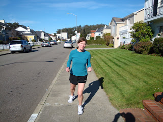
The lean to the left in this runner who just happens to be my wife Pat can be caused by a short leg.

When you watch someone walk, there may be a slight lean to one side which is very consistent. This is caused by many problems, including scoliosis and short leg syndrome. When evaluating for a possible short leg, look for this lean known as "limb dominance". Pat is demonstrating left side dominance. If you have received lifts for a short leg, the lifts should eliminate all or most of this lean. Never use lifts if the perscriber does not watch you walk and/or run with and without them documenting their success. Never use lifts if the lean is made worse by the lifts. If your symptoms seem to be getting worse with lift therapy, stop using lifts for a week to verify that the symptoms are being caused by the lifts. When they work, it is wonderful and life changing. When they do not work, search for a reason.

When you are walking along, do you ever notice you always drift to one side or the other? Do people who walk with you comment that you always bump them if they walk on your right or left side? Do they sometimes joke that you won't pass a sobriety test even when sober? If you walk with hard soled shoes on a hard surface, do you hear one side landing harder (greater sound)? These can all be clues of a short leg. With a short leg, you may lean to the long or the short side, but most adults lean to their long side.
dreamstime_m_13892607.jpg

In my practice I treat short leg for many reasons, but the most referrals I get are for hip and low back pain patients. Iliotibial band syndrome is also commonly caused by a short leg. When a patient is always injuring the same side, even though different areas, think short leg syndrome. Lifts to correct for the uneven hips and pelvis can allow the muscles to function equally on both sides of the body easing stress points and allowing muscle imbalances to correct. If you have any clues you may have a short leg, and you are having pain, perhaps lift therapy to correct for the short leg may be helpful for you.

Short Leg: Heel Lifts vs Full Length Lifts


The 9 common biomechanical categories used in the treatment of injuries are:
  1. Short Leg Syndrome (discussed here)
  2. Poor Shock Absorption
  3. Excessive Pronation
  4. Excessive Supination
  5. Tight Muscles
  6. Weak Muscles
  7. Miscellaneous Gait Abnormalities (ie Drop Foot, Polio, etc)
  8. Forefoot Abnormalities
  9. Combinations of the Above


The heel lift is the main method used in treatment of short legs and their problems. But, heel lifts are inherently unstable, and easy for patients to compensate for by simply bending the knee on that side. Once you bend the knee on the side you are trying to lift, you have lost the correction. Heel lifts, therefore, can make the foot/ankle more unstable by lifting your heel up in the shoe more, and they can make the knee more unstable by producing greater knee bend/flexion.
dreamstime_m_20520321.jpg
Doctors are always placing the lift onto an orthotic device. This is not advisable. First of all, it makes it hard to decide what symptoms are related to the lift, and what symptoms are related to the orthotic device, if pain increases with orthotic devices. Secondly, because of the instability mentioned above, the heel lift may negate the added stability produced by the orthotic device.

Golden Rule of Foot: Keep orthotic devices and lifts for short legs totally separate.

Thirdly, most of the lifts I have seen attached to orthotic devices are very hard for durability, but poor for impact shock absorption.


Full length lifts in the treatment of short leg syndrome are much more stable and produce better symptom relief. Full length lifts need to lift the heel and forefoot (metatarsal area) equally, then taper at the toes. This full length lift above, designed around a shoe insert template for W9 (women's size 9), is actually cut off at the toes to give room in the toe box area. Imagine all of the athletes who run up on the ball of their feet, but need a lift for their short leg, how could a heel lift do anything positive for their biomechanics and symptoms? At least 50% of the time most athletes are on the ball of the foot while exercising, so a full length lift seems ideal for the athletic population.

dreamstime_m_31830213.jpg

Now athletes on the other hand would love the lift to be only under the heel for convenience and shoe fit, but it is so much better to have it full length. Compromises are made all the time for lifts between ¼” and ½” (in even roomy athletic shoes or dress shoes with removable foot ends), typically the full lift needed is placed under the heel with ½ of the lift at the ball of the foot.


If you use full length lifts, make sure you make cuts into the material by the ball of the foot to help with smooth push off. This is especially true as you get up over 1/4 inch in lift. Material under the ball of the foot can make it hard to move through, so cutting to increase bend, along with beveling and thinning slightly can all help.
You can also see that the front edge of the full length lift is bevelled or skived to make a smoother transition forward. When I am using full length lifts, I always try to get feedback from the athletes on how easy it is to move through their foot.
In podiatry talk, I am trying to avoid Sagittal Plane Blockade.


You can see the cuts do not go through the sides to help with its durability, but they do go completely through from top to bottom.


Not really demonstrated well, but these cuts do improve the bend of the foot making it easier to move gently through to the toes. Many shoe repair stores have material to make a shoe lift.

Short Leg Treatment: Shoe Lifts

Most patients have a short leg, either structurally or functionally, or a combination of both. A structural short leg is true length difference of the bones, where a functional short leg may be caused by many factors including one arch lower than the other side. As long as one arch collapses more than the other side, the short leg syndrome exists.

Most people have one foot longer than the other, but the long foot may or may not be on the long leg. Remember you are taught to buy shoes always for the longer side (longer foot). If you wear out one shoe more than the other, either by observing the heel of the outersole or the footbed within the shoe, you can tell that one leg may be shorter. Orthopedists normally do not recommend treatment for leg length differences unless over 1/2". Podiatrists have observed that as little as a 1/16” to 1/8" difference in leg lengths can cause symptoms. By treating these small differences, and having patients report positive outcomes, leg length discrepancies are a vital part of care.

Treatment of leg length discrepancies is with various types of lifts under the short leg. The photo above shows a shoe with a full length external or outersole lift of 3/8". Due to the swelling in his foot, this patient could not tolerate any lifts within the shoe. Full length lifts, whether within the shoe or on the outer sole, are normally so much more stable than just heel lifts as previously discussed.

Most athletic shoes can accommodate up to 3/8" lifts. All patients should have a trial of lift therapy with shoe inserts with positive results before external shoe lifts are utilized. Most shoe repair shops can put on external lifts, but there may be one in your area that specializes. Ask around for referrals from local orthopedic or podiatry offices. The external lift must be tapered at the toes, and somewhat flexible at the ball of the foot, to allow the patient to walk smoothly from heel to toe. Any external shoe lift pushes the foot away from the bottom of the foot, so mechanically until we adapt to it, you must be careful walking on stairs (foot strike is earlier).

Golden Rule of Foot with lift therapy: Start Low, Go Slow. Normally, if the difference is 3/8" total, 1/8" lift is given for 2 weeks, then another 1/8" lift for 2 more weeks, then finally the full 3/8". As you go up in lift therapy, blame any new symptoms on the added lift, take out the additional lift until the new symptoms subside, then try again. Some patients are stuck for one reason or another at one level of lift. Their bodies will reject the higher amounts. There seems to be more stress on the body when the exact same lift is placed on the outsole, or as a midsole lift, as was originally used as an insert. It probably weighs slightly more, or affects the motion around heel strike more. To lessen this change, which may cause symptoms itself, place 1/2 of the overall lift in the opposite shoe as an insert initially. Two weeks later, take 1/2 of that away, then finally 2 weeks later take it all away so you are left with just the desired outsole lift. This eases the process dramatically, allowing the body to relax more in making this big change.

Understand the 3 Measurements taken on a Standing AP Pelvic Xray
Standing AP Pelvis Xray with shoes and orthotic devices

Look at the heights at the Acetabulum, Sacral Base, and the Iliac Crests

As you review this x ray, you will see that the the left hip at the acetabulum (hip joint) is higher than the right. This is the true leg length difference if the foot is in its neutral subtalar position (why it is best to take this xray with stable shoes and stable orthotic devices in the shoes). The symbol marked UPRIGHT means standing and is on the left side. The base of the spine where L5 vertebrae rests on the sacrum, also called the sacral base, drops to the right side. Many feel that getting a level sacral base is more important than correcting the hip height difference.The highest point on the iliac crest, not even seen on the left since it is higher than the right side, is a summation of the pelvic difference. When you look at many points on the pelvis, comparing right to left, you will see how the left is higher all around. However, one of the major problems we face is one of trying to take xrays only when crucial (due to the radiation). I would love to x ray after every change I make, but I must be conservative due to the radiation exposure.

Common X Ray Findings Example:
  • Hip Height at Acetabulum 10 mm short right (TRUE STRUCTURAL)
  • Iliac Crest 17 mm short right (further pelvic and Sacroiliac joint collapse)
  • Sacral Base 13 mm short right (amount spine needs for leveling)
With these findings, it is easy to start with 1/8th inch lift (3.3mm) for 2 weeks, another 1/8th inch for 2 weeks, and then 3rd 1/8th inch lift for the final 2 weeks. I start with tie-on shoes that take the full 3/8th of an inch. I would then have the patient wear this amount for two months to get use to them. Some time with a physical therapist to work out the predictable muscle soreness that will ensue would be great during this time. After the 2 months, if there is still limb dominance, still some symptoms, then I would go up another 1/8th to correct for the sacral base (the extra 3 mm the xrays showed). Of  course, many stop the correction at lower levels if the symptoms are resolved. Patients can fight you a lot during lift therapy since they do not want lifts. Who would?? It is important to stay focused, and the xrays really help in this regard. The xrays tell us what is the short side, and by how much. Treatment can be gradual, but complete correction of a short leg is usually obtainable. Watching walking and standing habits you can see these gentle habitual leans (dominance) that can be a sign of short leg syndrome.

dreamstime_m_30439949.jpg
Gentle lean to the left of the girl standing noted

Sunday, December 21, 2014

Inverted Orthotic Technique to Correct Flat Feet in Children

http://synapse.koreamed.org/Synapse/Data/PDFData/1041ARM/arm-38-369.pdf

     I am so proud of this article published by researchers in Korea. It substantiates a claim that the Inverted Orthotic Technique, which I developed throughout the last 30 years, actually helps children with flatfeet improve (called pes planus). I have used the technique on 100s of children, including my own two children, starting at 3 for one, and 1 and a half for the other more pronated child. If your children have flatfeet, have a podiatrist evaluate if they should have orthotic devices and when. I have tended to never treat my family as a general principle, but in this case, I have felt the most qualified.

Monday, December 15, 2014

Complex Regional Pain Syndrome: Email Advice

Dear Dr.blake
Hope you are doing well .
Just wanted to update you regarding the progress of my algodystrophy (that struke me after my sesamoiditis ) Dr Blake: Algodystrophy is another name for complex regional pain syndrome.
As this might help other people who are in pain or experienced RSD symptoms.

-          I have followed your advice and went looking for calmare therapy – couldn’t find any around since I live In a small country – I found one center who have a PBK “Italian electro machine”
The owner claims that he have amended it’s program to do the effect of calmare/ since I had no choice I tried it /
To my surprise it worked wonderfully. And after only 6 sessions  . my pain was 50 % less . now I have high hopes , I think by Christmas my aldodystrophy will be gone  "6 month after the discovery of the RSD”
So I definitely recommend calmare or alternative calmare ,  since I have tried acupuncture and other  it helped only at first.   Dr Blake: Calmare works permanently on some and some seem to need booster sessions occasionally. Calmare is an electrical current and does not involve anything invasive, like IVs or injections. 

-          My main problem is that after I heal from the main full leg pain I would still have issues with my fingers and sesamoid bones ,  because of the non bearing issue – what shall I do or start doing to improve mineralization in
That area. And as per your  experience how much time it would take to get back to normal in that area after RSD is gone. I am seriously desperate to go back to normal and willing to do anything required.
Dr Blake's comment: This patients has had very bad disuse atrophy of the bones from months of non weight bearing and inactivity. The treatment needs to be directed towards gradual weight bearing. There is no time lines that I know. Set benchmarks on what you can do this month, and reset them in next month. By gradually increasing walking, biking, elliptical, core work, etc. the strength will come back, but never at the pace you want. 

-          I have another issue , with orthotics ,  I have tried a dozen with a collection of shoes . and paid of fortune on these . still I have a problem that when I stand more than 20 minutes in these ,  I feel like I have been standing all day, and
I have to sit for a while before I can walk pain free. Also walking for more than 15 minutes give the same feeling.
I got some weird pain till the point that I thought I have RSD in the second leg . but I don’t /
I know orthotics are not supposed to be 100% comftable and that they changed our gait .   but this is really frustrating and I feel it in both legs , if I have to wear orthotics for a year or so , I really need to find a solution.
An orthopedic doctor told me that my calfs became weak and there’s too much pressure on the feets and heels= he said that  after a year of wearing orthotics my gait changed and my muscles got weaker and that I need to do stationary biking – I cannot do biking now and waiting for a month or so when RSD is fully gone.
Is that kind of pain and discomfort normal when wearing orthotics ,  what can I do to reduce it ???
Dr Blake's comment: All you can expect orthotic devices to do is protect the joint that was injured. Your symptoms seem to be bone fatigue and muscle fatigue and nerve irritability. You seem to be on the right path to restrengthen. Find the most comfortable orthotic you have and experiment with more padding, more arch support, more dancer's pads, etc. See if anything mechanical really improves the tissue threshold you are experiencing now. 
-          Regarding Vitamins .  the doctor told me to reduce smoking and it helped . he gave me raw honey since he said some new researches revealed that RSD is linked to a deficit in the immune system.
And I am taking multivitamins with extra calcium and magnesium.  For how long do you think I should continue taking vitamins ? ? is it ok to take for long months or years or this could be harmful.
Dr Blake's comment: I think that you should sit down with a registered nutritionist and attempt a game plan to reduce inflammation, improve nerve and bone health, and strengthen your immune system. 


Sorry for the long email.

I thank you a lot for your support and wish you happy holidays . Jesus bless for being such a hope for many people around.
Dr Blake's comment: Thank you, I have been so blessed by God that it is hard to ask for anything else. Merry Christmas to you. 

Friday, December 5, 2014

Accessory Navicular: Email Advice

Dr. Blake,

I have been intently searching the Web for some insight on accessory navicular and you seem like a Dr. with a solid understanding of the disorder.  I don't know whether or not you are willing to give me some advice but I thought I might ask???
Dr Blake's comment: The accessory navicular only occurs in 10% or less of the population. If you look at the illustration below, look first at the heel bone. As you move ahead towards the arch, you will see a small bone (accessory navicular) under a bigger bone (navicular). This is where the powerful posterior tibial tendon attaches and helps support the arch.The accessory navicular makes this attachment weak. 


My son is very athletic and loves to play basketball. 


 He was born with nice arched feet.  Last spring he was playing basketball with slip-on sandles and one of his bigger friends stepped on his foot injuring him.  He came home limping but was able to continue playing basketball.  While in a game a few days later something happened to this same foot, it rolled.  We backed off on it but it continued to hurt so we took him to Childrens ER where the Dr. diagnosed my son with accessory navicular.  He explained to some degree what we were dealing with.  The injury occurred in April 2013.  The Dr. gave him some arch supports and along with PT and Dexamethasone, and the Dr. said in 6 weeks he said he could start playing ball again.  We held out letting him play full B-ball until late June 2013.  After a two day tournament he said it was seriously bothering him again.  Needless to say, he started PT again and Dex.  In August we went to a specialist and he gave my son custom orthotics.  We leave the orthotics in his everyday shoes.  We bought him new basketball shoes and put some OTC sports orthotics in them with a high arch.  He is 13 now and playing school ball, he plays alot.
Dr Blake's comment: For playing he should be in the most supportive insert (based on his feel) and be taping. Try one of the taping techniques on my blog, or go to www.supportthefoot.com and order small size pack from them. Very supportive and can last a few days. 

His name is Luke and prior to his injury, never complained once about his feet.  He can run two miles right now with zero problems.
Questions??
Do you think he is going to be fine or do you think this issue is going to keep surfacing? Dr Blake's comment: Unsure, but it is not a predictable sign that surgery is in your future. 
He is very inflexible, he cant come close to touching his toes, he used to be able to.  Do you think this is because of his condition??
Dr Blake's comment: Probably not, one growth spurt can do that to you. But, very important, unless it causes pain, to stretch out the achilles well on a daily basis. See the upper right stretch.


He walks like an old man sometimes, sometimes walking down the steps very slowly, especially after a day of playing hard.  Do you think this is because of his "AN"? Dr Blake's comment: Unsure, may just have played hard.
Other times he looks great on his feet. Dr Blake: When you have a really bad problem, you never look great until it is fixed. Kids are always hard to read. 

Do you think the Kinesiotape will help, if so how should I wrap it??

http://www.drblakeshealingsole.com/2014/04/arch-taping-with-kinesiotape-kt.html

What type of strengthening should he be doing?

http://www.drblakeshealingsole.com/search/label/Foot%20and%20Ankle%20Strengthening%20Video%20Playlist

 Dr Blake: Find out which ones he can and can not do without pain. Pain actually shuts down the muscle and makes you weaker. I hope this helps some. Rich

Thanks for any input you may be willing give!!!  I would very much appreciate your input.

He used to run around all the time jumping everywhere now I really dont see him jump around that much, unless hes playing ball.

Biomechanics Medical History

Taking a Good Biomechanics History
dreamstime_s_16250464.jpg

    This is where it all begins in the doctor/patient or therapist/patient relationship. The time spent here discussing the historical facts of an injury or pain syndrome, and important contributing factors, can be vital in the success or failure of treatment. Why is it so vital? Follow up visits work off the success or failure of the treatment plan set on that first visit (it is why I am anal with staff to allow that patient ample time, and allow me to see them on time). If the information collected is inadequate, the entire sequence of events following may be subpar. Please review Chapter 3 on History Taking now before we go further.

    The biomechanics history related to injuries is looking for patterns or facts that can cause injuries to occur. Here are some of the many questions that normally get asked, or at least you should add to your thoughts prior to seeing a doctor or therapist. These include:

  1. Do you know if you have a short leg?
  2. Do you believe you have weak or tight muscles in general, or around the injured part?
  3. Do you have loose ligaments in general?
  4. Are you right or left handed?
  5. When you were a child did you have to wear braces or shoe inserts?
  6. Have you ever been prescribed shoe inserts?
  7. Have people told you that you walk or run funny?
  8. What has your history been of overuse injuries (non traumatic)?
  9. Have all or most of your injuries been to one side of your body?
  10. Do you have high arches, flat feet, bow legs, knock knees, bunions, hammertoes, or other abnormalities?
  11. Do you have any arthritis from your hips downward and where?
  12. Do you feel unstable in any joints?

    A skilled practitioner knows the relevancy of the answers to your problem. The answers will help point the course of treatment in the right direction. I sure hope it helps you.


dreamstime_m_41933283.jpg

Wednesday, December 3, 2014

Nerve Pain and Gait Changes with Drop Foot: Email Advice

Hello Dr. Blake,

I recently discovered your blog and certainly appreciate the expertise you share.  I see that you are aiming for the Camino de Santiago.  I would like to travel part of the camino but am struggling with my current right foot condition.  Having grown up in Madrid, Spain, I believe that you will enjoy the journey.  I am 65 years old and recently retired!  I am 5'4 and weight 120 lbs.  Other than the spine and leg issues, in good health.

In July 2013, I was diagnosed with L5 radiculopathy with right foot drop (significant).  This was very painful and the pain was mainly below the knee.  MRI did not indicate the L5 radiculopathy, only an EMG. (Dr Blake: 1 problem with low back MRIs is that they are done in spine neutral, most stable position), and not when the spine is stressed so problems with bulging discs can be missed).   After 60 PT sessions, including traction and short term use of an off the shelf AFO (which caused medial pain and per the physiatrist plantar fasciitis), and the passage of time, I have recovered much of the foot control without any surgery.  What remains has caused havoc in my life due to medial foot pain.  I have also had an orthotic with a cork base which was adjusted many times by an Orthotics Center but which I believe contributed to subsequent problems due to a tendency to "roll" the foot due to the weak everters.  I have used KT (kinesiotape) in many ways to support the foot.  More recently an MRI showed a split longitudinal tear of the Peroneal Brevis and tendinosis of the Peroneal Longus (Dr Blake: these tendons can look like this in normal ankles, so difficult to take too serious unless surrounded by major inflammation).  This occurred after initiating additional PT.  The PT indicates that I have a very weak Posterior Tibialis and is also making me a new orthotic that is to support the believed weaknesses better than the prior.  I am awaiting the new one shortly.

I recently had a gait analysis  at a Motion Analysis Center in Michigan, and the report indicates:
*** Dr Blake's notes in red
Mild R forefoot inversion in stance and swing (perhaps anterior tibial spasm)
Mildly decreased R ankle dorsiflexion in early swing (some foot drop)
Minimally decreased ankle dorsiflexion in early midstance bilaterally (antalgic stiff gait)
Mild forefoot adduction in stance bilaterally with R slightly worse than L (perhaps anterior tibial spasm)
Mildly decreased ankle plantarflexion moment in stance bilaterally (stiff not propulsive gait)
Moderately decreased ankle and hip power generation in pre-swing bilaterally
Pelvic rotation pattern reversed versus normal with protraction rather than retraction in midstance.

A fine wire EMG indicated that the TP was active 15-30% of the time when it would normally be active and only during the late mid to terminal stance (so would say 50% loss of Post Tib function)

You would not think that something that is mild/minimal would cause so much pain in the medial foot.  Certainly the lateral foot PL and PB issue causes some discomfort but that is not the main cause of pain.

Recently was referred to another physician that uses ultrasound of the tendons and he said that arthritis of the first metatarsal may be the primary issue and gave me a steroid injection which for a couple of hours helped a bit but then the joint pain was significant.  The orthopedic surgeon that referred me to his colleague for the ultrasound wondered if the problem may be the Anterior tibialis insertion in the Medial Cuneiform.  His colleague did not. (I would have to say Anterior Tibial spasm compensating for weak PT tendon and weak extensors--the ones that causes the drop foot---would be my first choice)

Given the time that has elapsed what might be fixable?  I am willing to have a custom AFO too.  Icing does not usually help.  Ibuprofen minimally helps.  I cannot take Neurontin due to side effects. 

So....the physicians are perplexed about what to do to help me.  I am awaiting the new orthotics which may or may not help.  I am interested in your thoughts/questions to help sort this out more. 

Greatfully,

Dr Blake's comment: I am assuming that the MRI did not show a tear in the Anterior Tibial Tendon or Arthritis near the Medial Cuneiform. Have them check if the Anterior Tibial is in spasm. Local anesthetic blocks into the muscle can break the spasm, and biotox is also being used. If it is truly the anterior tibial spasm that is dorsiflexing the ankle and holding up the medial arch, then an AFO should be used until you gradually regain full strength of the ankle, and normal heel to toe motion. Let me know if you have other questions. 


Tips to Avoid Cast/Immobilization Problems

Tips to Avoid Cast/Immobilization Problems

Injuries can bring with them some form of casting to protect the area for a long enough time that the injured part has a great chance to heal. But as healing occurs to one area, other areas can be negatively impacted. The forms of casting available include permanent (plaster or fiberglass-type) and removable. All forms of casting cause a syndrome affectionately called “Cast Rot” or “Cast Disease”. This syndrome includes:

• Muscle/Tendon Atrophy or Weakness
• Muscle/Tendon Stiffness or Loss of Flexibility
• Proprioceptive Nerve Problems with Loss of Position Sense
• Cardiovascular Loss
• Weight Gain due to lessened activity
• Hip/Back Problems when the foot/ankle is immobilized due to height difference from cast side to good side

dreamstime_m_18724953.jpg
The permanent cast by far produces the most problems, but even the removable casts can be very destructive. In the rush of reviewing the x rays or MRIs, making the decision to cast, and orchestrating that the proper cast is applied, many simple suggestions are forgotten that can minimize some of the effects of cast disease. If you have a foot/ankle cast, or boot make sure the opposite foot is raised up evenly. A product called EvenUp® can be applied to the shoe on the opposite side to keep the knees, hips, pelvis, and spine level (as demonstrated in the photo above). Find out what cardio you can do to keep the heart/lungs strong (and remember the HDLs) and contain inevitable weight gain. Some walking is normally allowed with weight bearing casts as the soreness resolves, but stationary bikes on a daily basis can keep leg strength and adequate cardio.

dreamstime_m_33772254.jpg
Normally you can do pain free isometric strengthening exercises within the cast, but a physical therapist will have to show you how to do these. Normally, push painfree into the cast in all 4 directions, hold for 6 seconds, and repeat 10 times. Do these isometrics 3 times daily. Sometimes, it is appropriate to order a muscle stimulator. This can even be placed inside a permanent cast to begin strengthening as soon as possible. This is normally ordered and applied at the 2 week cast change.
dreamstime_m_28314408.jpg
As soon as the cast comes off, you may not be able to increase cardio, but you should be able to increase strength, flexibility, range of motion, and proprioception. Find out what exercise you can do as quickly as possible without risking harm. Keep pushing the doctor and/or therapist to move your rehabilitation along. A prescription for 3 physical therapy sessions (normally once every 2 weeks) can progressively build a home exercise program that you do daily.
Golden Rule of Foot: For every day you are in a cast,it takes 2 days to get back to normal. Your job, if you choose to accept it, is to move the rehabilitation along as quick as possible, without causing increased pain. Pain causes swelling, which causes more pain, which causes more swelling, and the cycle spins out of control. Be your own advocate, ask questions, make sure speedy rehabilitation is part of all those involved mindset for you. Tell them you want to soak to reduce swelling, you want to do exercises for strength, flexibility, range of motion, and proprioception. Tell them you need to get safe but effective cardio as soon as possible. Keep it moving! Another Golden Rule of Foot!!