Calcaneal (Heel Bone) Stress Fractures: A
Cause of Significant Persistent Heel
Pain
By Richard
L Blake, DPM
Heel stress fractures present the same way as plantar fascial tears. They present with swelling, typically an acute onset, and pain level in the 4-6 range or more. However, unlike plantar fascial tears, they may develop slowly probably progressing from a bone bruise, to stress reaction, and finally stress fracture. They do not show up on x ray normally, making an MRI or bone scan typically needed to confirm. Like plantar fascial tears, if this is suspected, and getting test confirmation is difficult to impossible, it is important to treat it as if it was a stress fracture. You do not want a calcaneal stress fracture to develop into a full fracture (typically needing surgery with some permanent disability possible). If you squeeze the heel from both sides, and you (the patient) is very sore compared to the other side, you may have a stress fracture. If you walk on your heels only for 3-4 steps, and you have excruciating pain, you either have a plantar heel bursitis or calcaneal stress fracture.
The top 10 treatments
for calcaneal stress fractures:
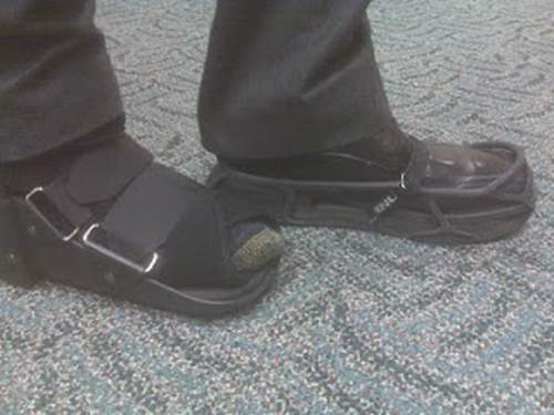
1. 3 months removable boot and EvenUp on the other side (and many times the heel bone has to be floated for off weighting with 1/2 adhesive felt under the midfoot and forefoot only))
2. 1500 mg calcium and 1000 units Vit-D3 daily
3. Bone density test if any question on why heel
broke (did not make sense?)
4. Vit-D3 level if any question on why heel broke (or if your dietary intake is low, and you do not get much sun exposure without sunscreen). This is especially true when the stress fracture occurs in the winter months)
5. Custom or OTC orthotic device to produce the effect of a soft
heel and weight transfer into arch
6. Ice pack 2x/day
7. Contrast bath each evening
8. Activity modification to maintain cardio
9. No NSAIDs like advil or aleve (slows bone healing)
10. Exogen bone stimulator for 9 months (if the
diagnosis is confirmed by MRI as x-rays are not great for stress fractures)
Patient
presents with swelling under the heel bone. There is pain produced on side to
side compression of the heel bone during physical examination. X-rays normally
are inconclusive. The patient does not have to have a story of landing hard on
the heel. Onset of pain normally occurs over a short time (acutely), whereas
plantar fasciitis (more commonly a cause of heel pain) has a typically gradual
onset of the pain, worsening slowly over a month or so. The typical
differential diagnosis with significant heel pain with swelling is calcaneal
stress fracture or plantar fascial tear, with some arthritic conditions much
more rare.
An
MRI is the conclusive test. It is important to note how close the stress lines
are to the subtalar joint. The closer to the subtalar joint, the more
consideration of non weight bearing 8 weeks of permanent casting (yes, a real
cast). This is totally devastating to a patient, so avoid when possible. The
following are 4 MRI’s for patients with heel pain, each with different
findings.
 |
|
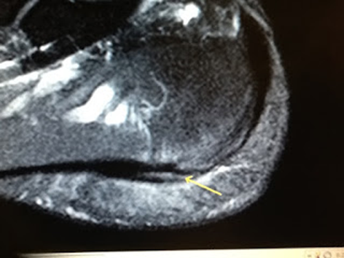
This MRI showed the bone
swelling above the bottom of the heel bone due to a tear in the plantar
fascia. You can see the intense swelling above and below the plantar fascia.
This is not the pattern of swelling of a calcaneal stress fracture. A small
blood vessel is seen running through the heel bone which can look like a
stress fracture. If it was there would have been reactive bone changes around it
eliminating that nice tortuous pattern. |
 |
|
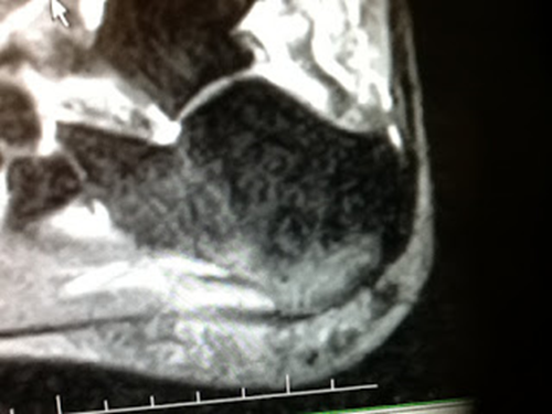
This is a tremendous
bone reaction from a calcaneal (heel bone) stress fracture that runs from the
bottom to the top of the heel to the subtalar joint. A permanent non weight
bearing cast for 4-8 weeks could be easily recommended to protect the joint.
This particular patient would have mentally lost it, so I did treat this with
a removable walking boot. She has done well, but did take longer than normal.
|
 |
|
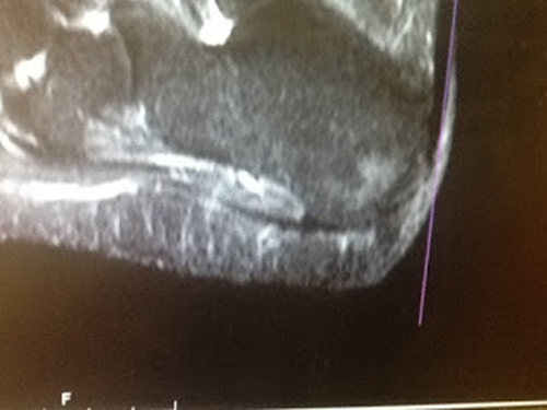
Same patient from just
above is 3 months into her treatment, still very sore, with still bone
swelling within the heel bone. As long as there is bone swelling, there will
be pain (like the pain you get from a sinus headache, although you never have
to walk with full body weight on your sinuses). I never created a good pain
free environment for multiple reasons, so the typical 3 months of
immobilization actually lasted 6. She was however able to do intense spin
classes and swim without problems during this time. We consciously as a
physician and patient team, traded early function for a potentially longer
rehabilitation period. |
 |
|
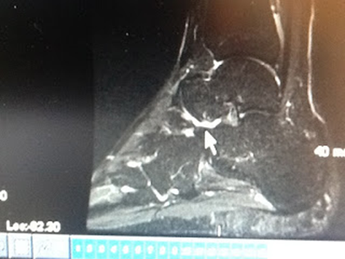
Normal heel bone with organized
blood vessels. |
Once the diagnosis is made, here is a checklist of
events that should happen:
- Questions should be asked about bone density issues,
dietary habits, activity levels leading to overuse, selection of shoe
gear, and past history of fractures.
2.The patient
should be fitted for a removable walking boot, unless concern that the
fracture goes too close to the subtalar joint. If the fracture is deemed needing non-weight bearing, a permanent cast is normally used for 4 to 8 weeks. I use a
1/2 inch accommodative pad to float the heel of the walking boot, and tend to
use a below the knee cast over a shorter one. An EvenUp is used on the other
shoe.
3.Over the first 2
weeks post diagnosis, you strive to create a pain free environment. The ease or
difficulty in creating this pain free environment is an important clue on how
serious the problem is. The average patient needs to be in the removable cast
for 3 or more months once the pain free status is attained.
4. Activity modification
is crucial at this time. Bike and swimming are commonly used to maintain
cardio, especially if a removable boot is used. Floor exercises for strength
and flexibility are recommended. Pilates is a great source of these exercises.
5.Sole, PowerStep, or PureStride OTC
orthotics are used within the cast (and later in the shoe gear) to produce
heel padding and weight transfer into the arch.
6.Contrast baths
once or twice daily are vital at reducing heel bone edema (swelling). Swelling
within the bone should be minimized since it actually can reduce the normal
blood flow important for healing. This can slow healing.
7. A Bone Stimulator for 6 to 9 months is used. I actually stop 2 months after full activity is resumed. I use Exogen ultrasound for this, but there are other good stimulators. For insurance, since there are no fracture gaps in a calcaneal stress fractures, many will not cover.
8. The Primary
Care Doc should discuss all the factors that affect bone healing including the
right amounts of calcium, Vit D3, and other minerals. With bone injuries, I
have the patients minimize their use of NSAIDs (like advil, etc).
9. Monthly return
visits can be scheduled for a while to monitor the progress and make changes.
 |
|
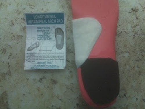
Sole OTC inserts with
extra cushion in heel and extra Hapad arch support to transfer weight into
the heel. |
10.
One month after the diagnosis, the patient is normally casted for custom
fitting soft orthotics. I use the Hannaford technique, but most professional
orthotic labs have their versions that can/are similar. These are dispensed in
1-4 weeks depending on the need to see that patient (if the pain free
environment is established already, waiting 4 weeks to dispense the new
orthotic devices is probably fine).
 |
|
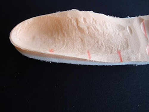
This
shows the memory foam of a Hannaford soft based custom orthotic device. |
11.
One month later, normally now 2 months post diagnosis, physical therapy can be
started to decrease inflammation and work on the damaging aspects of casting:
stiffness, weakness, loss of proprioception (balance), and sometimes nerve
hypersensitivity. Physical therapy can be helpful until you are back to full
activity, probably 3-6 months. Most of the time physical therapy can be
effective at 1-2 times per week.
 |
|
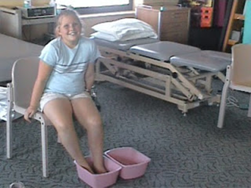
Patient in physical
therapy doing contrast bathing to reduce bone swelling and its resultant
pain. |
12.
Three months post diagnosis should mean that the patient has been pain-free for
almost exactly 3 months with all of the above treatments. If it was tough to
get the pain level under control, then this landmark may take much longer. It
seems that the patient can successfully wean off the removable boot after being
relatively pain free for 3 months, no matter how long that takes. To
successfully wean off of the boot means that you can not have more pain out of
the boot than in the boot. The removable boot or cast (I use those phrases to
mean the same thing) is initially weaned off by keeping it on at work, and
gradually adding more time out of the boot at home or doing errands. When you
are completely weaned out of the boot for home, gradually spend less time at
work. During this time there can be no increase in pain, you should ice 2 or 3
times a day extra (ice pack 15 minutes to the bottom of the heel), and the
whole process can take 4 to 6 weeks. During this time always have the boot with
you!! You never know when you will need it. Once you are out of the boot full
time, you can gradually increase your activity.
No comments:
Post a Comment
Thank you very much for leaving a comment. Due to my time restraints, some comments may not be answered.I will answer questions that I feel will help the community as a whole.. I can only answer medical questions in a general form. No specific answers can be given. Please consult a podiatrist, therapist, orthopedist, or sports medicine physician in your area for specific questions.