Welcome to the Podiatry Blog of Dr Richard Blake of San Francisco. I hope the pages can help you learn about caring for foot injuries, or help you with your own injury.
Hintermann published a paper in 1996 about a clinical test to help him decide whether a patient needed surgery for posterior tibial tendon dysfunction. It was based on the fact that with the patient standing, when the heel is inverted (or the leg externally rotated), only in the 21 patients that had posterior tibial disease (not necessarily ruptures) was this test positive for leaving the first metatarsal off the ground. It is now known more as the First Metatarsal Raise Test. When I read the article many thoughts crossed my mind, and I need to do this test some, but I need other podiatrists to give me feedback on their successes and failures. So, what bothered me about this test? The things that bother me are:
100% of the patients were positive even though the surgical findings were all over the place (from tendinitis only to complete ruptures)
100% of the patients without post tibial tendon disease were negative, but they do not go into any of these patients (allow they implied the test was being done over 4 years)
They made no reference to what type of orthotics were being used preop to avoid surgery and whether the tendon being strengthened thoroughly before (there was no mention about posterior tibial strength at all)
They seem to have no knowledge of deformities like rearfoot varus, rear foot valgus, forefoot valgus and forefoot varus. Any of these common deformities would greatly affect this test.
In most of my patients with PTTD, with 10 degrees of heel eversion, and 10 degrees of positional forefoot supinates, when I put the patient into heel varus the first metatarsal is going to be way off of the ground. This does not mean I need to do anything but rehabilitate them.
I hope you are doing well and staying healthy during these crazy times!
I have been thinking of writing you for quite a while, but my question is quite lengthy.
I have attached some x-rays and photos to this email, but I also have a CT cat scan
and an MRI of my foot if having those would be better.
Dr. Blake's comment: Yes, send those to Dr. Rich Blake, 90 Beachmont Drive,
San Francisco, CA, 94132 to see if those are revealing.
I have developed a hallux claw toe in my right foot after I had a bunionectomy
and tibial sesamoidectomy in 2014 and I am wondering
if I should get surgery to prevent a joint fusion surgery in the future.
My toe does not bother me terribly now. Every other day I can go on a 1 mile walk,
with minimal discomfort. However, I have noticed that as time has gone on
it has become progressively more sensitive
(able to walk less and less mileage without the next day hurting)
and I want to maintain a relatively active life as I am 33 and a mom.
My right big toe does not really touch the ground, and my other four toes
on the right foot are starting to develop hammertoes.
My left foot also has hammertoes so I know I am prone to them,
but they are still relatively mild on both feet.
I have been recommended two different surgical techniques to prevent further problems
down the line with my foot, and am wondering whether you think
1) surgery is a good idea, and 2) if so, which surgery you think I should opt for.
History:
Initial Breaking of Tibial Sesamoid with subsequent Surgery: I have high-arched feet,
and have worn orthotics since age 13 or 14.
I broke my tibial sesamoid after wearing heels to a party in 2013.
It was described as a freak accident (as I guess most bone breaks are!)
but people had always remarked at how high my arches were,
so I guess the pressure was too much. 7 months later
I was told there was no healing and it was recommended I have a tibial sesamoidectomy
as well as a bunion surgery (chevron osteotomy),
as I already had a bunion but the broken sesamoid was accelerating its progress.
I wish I had found your blog back then, as it was described as a relatively easy surgery
with minimal complications. After the surgery I went to physical therapy.
It was always hard for the physical therapist to get my big toe to touch the ground even then.
A year after the surgery I noticed my right big toe no longer touched the floor when barefoot.
As long as I wore orthotics in shoes I was pain-free and comfortable
and could go about my daily life. Walking barefoot was uncomfortable
because the ball of foot was pushed into the floor without weight bearing from the big toe.
Dr. Blake's comment: This is a common problem with the tibial sesamoid removal which weakens the short flexor under the joint. This enables the tendon on the top of the toe to win, called the extensor.This dynamic imbalance pulls the toe up starting the hammertoe. With the pes cavus or high arched foot, you probably even before surgery had a tighter extensor ready to pull the toe up. With this being said,
it does not explain why the PT could not get the toe down which let this get out of hand.
Most Recent Setback: In May 2019 I wore a pair of heels on date night
and walked about 10 minutes on the cement sidewalks. (No heels allowed in my closet again!)
The next day my fourth toe and especially my big toe were very inflamed,
and it took 5 months of me wearing a boot and daily icing in order for the symptoms
to calm down. This is when I found your blog. During those 5 months
I visited multiple podiatrists and two orthopedic surgeons to try and figure out
why it was taking so long to heal. It was determined that I stretched the collateral ligaments
in my stiff big toe. The second orthopedic surgeon had a nerve-conduction ordered
to ensure I did not have charcot-marie-tooth disease, due to my high arches,
raised toe, hammertoes and slight atrophy of right-side calf muscles.
Luckily it was determined I do not have the condition, but both orthopedic surgeons
commented on how raised my big toe was, and were concerned about how my foot
would handle such biomechanical stresses for the duration of my lifetime.
Both of them suggested some surgical techniques to prevent the further deterioration of my foot.
I know you are not a proponent of “preventative surgery”, but I am wondering
what you recommend in my case.
Dr. Blake's comment: All we know right now in this correspondence is that you have a very sensitive
big toe joint. I think everyone, except a few surgeons, would agree that an operated on joint, is and will
be forever a weaker joint. When you do hip surgery, and slightly less with knee surgery, you can
establish great stability on the surgical site with the surrounding muscles. You
have great opportunity to really tone them up. The foot has some crucial
muscles to help, but as podiatrists we mainly have to rely on shoes, orthotics, accommodations, taping,
etc, and physical therapy to breakdown scar tissue, remove inflammation, and stretch and
strengthen the muscles up the leg to the hip.
Current Pain and Issues:
1) My current symptoms are that my toe is quite stiff at the MTP joint,
and semi-rigid at the IP joint. I have good range of motion with the toe going upwards
but pretty limited motion when trying to force my toe down.
The bottom of my big toe is sore (scale of 2 out of 10) if I go on a 1 mile walk or more.
If I go on these walks for more than a couple of days I have to take the next day off.
Furthermore, the side of the top of my big toe gets sensitive too if I go on long walks
or walk barefoot on hardwood floors. I have noticed this is because my big toe
actually turns into the rest of my foot, in addition to the claw toe.
It feels like the bone is pushing out? This varus has been increasing over time.
Dr. Blake's comment: So, if you were in my office I would have you practice and get really good at
spica taping to hold the toe down for the next 8 weeks. Patients usually can advance from the easier KT
or Rocktape to the 3M Nexcare Waterproof tape. You should have a PT over the next month to year
(when corona quarantine is lifted) measure the amount of plantar flexion of the big toe joint which
Welcome to my Google Document that I shared with my Podiatry Students at the California School of Podiatric Medicine Samuel Merritt University Oakland, California. This was my first Zoom teaching, so I over prepared, and dropped everything else I was doing last week, but I think it went well. I will be putting the answers to questions on this on request of student doctor Diksha Mohapatra by April 17th, 2020. The videos are great to watch if you want to learn about gait evaluation. Rich Biomechanics Workshop Outline (4/10/2020 1-3 pm)
Introduction of Students and Dr. Rich Blake
Allison Ma
Amy Lau
Andrew Rasiah
Diksha Mohapatra
Maksim Rukhalskiy
Shane Hall
Usman Khokhar
Yona Mizrahi
2 Classes from Dr. Rich Blake Gait Evaluation 4/10/2020
Orthotic Devices on 4/24/2020
(we will start next session with the discussion of the assignment below on Golden Rules)
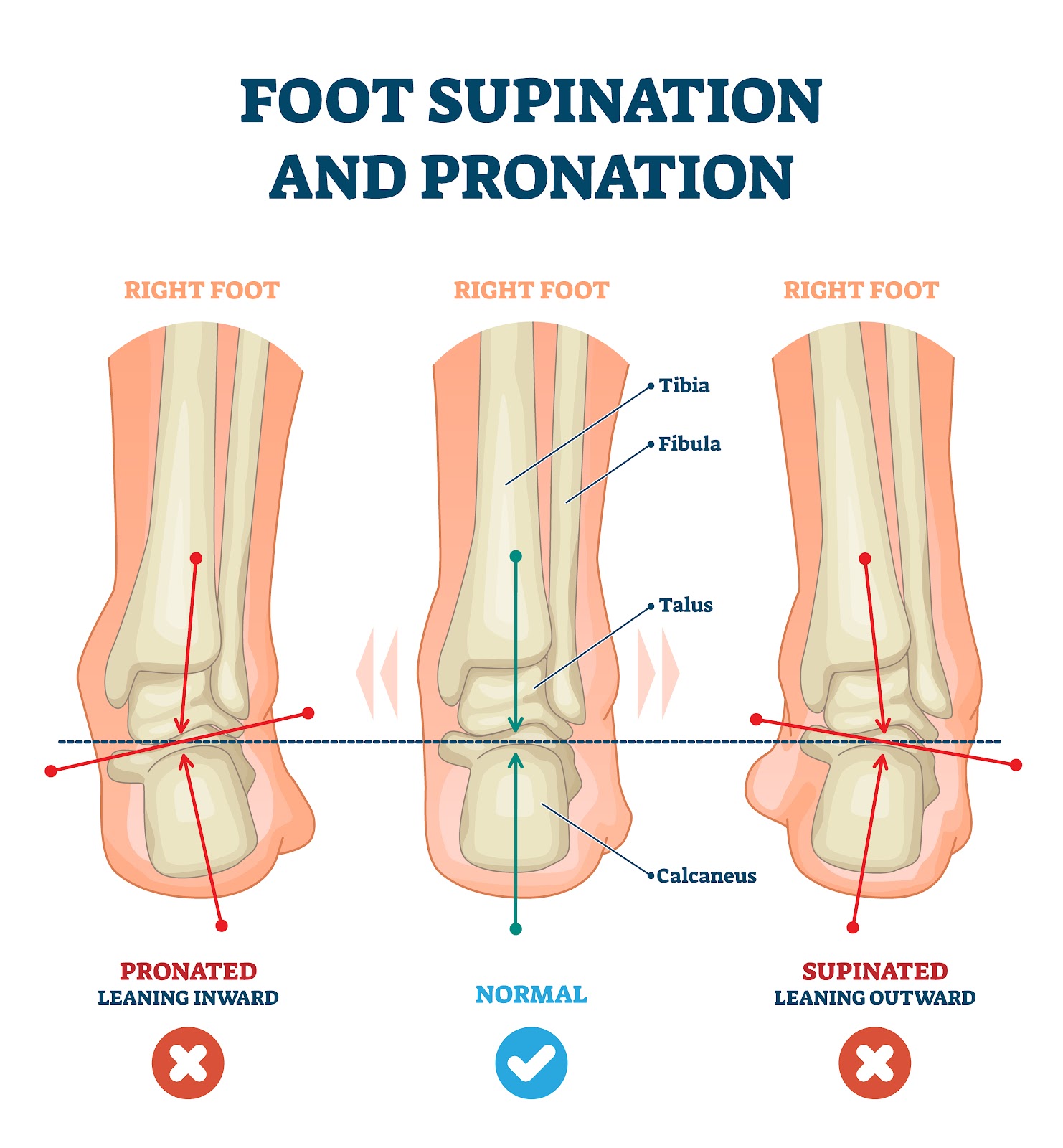
I hope to teach you that Gait Evaluation is much more than pronation or supination of the foot. Look at the whole body. We use gait evaluation as a clue to find out what is wrong with a patient, and how to help them.
Right Foot
Some of your patients will like this video, so you should know how to treat them and we will go over orthotic devices next session.
Allison: What are the 2 separate parts of gait called?
Amy: What are the 5 components of stance phase?
Andrew: What are the 3 components of the swing phase?
Diksha: With what you know of the contact phase as the ankle is plantar flexing, what is the heel doing in the frontal plane and the knee doing in the transverse plane?
Maksim: How does the patient prevent foot slap gait with drop foot?
The common findings in gait that you as a podiatrist will see in regular practice is less pathological and more subtle, but still very important. Looking to see how to help each patient mechanically.
Ability or Inability to Walk (First and Foremost)
Structural deformities like pes planus, pes cavus, bow legs, knock knees, genu valgum, etc.
Signs of Asymmetry
Signs of Pronation
Signs of Supination
Signs of Limb Length Discrepancy
Signs of Tight Muscles/Tendons
Signs of Weak Muscles
Signs of Poor Shock Absorption
These are all problems that we can make mechanical changes to help
Maksim: What would be a sign of pronation?
Shane: What would be a sign of supination?
Usman: What would be a sign of short leg syndrome?
Yona: What would be a sign of tight muscles?
Allison: What would be a sign of weak external hip rotators?
Amy: What would be a sign of weak achilles tendons?
Andrew: What would be a sign of weak peroneal tendons?
Diksha: What would be a sign or weak posterior tibial tendon?
Maksim: What would sign of poor shock absorption?
Below is a video with a more podiatric approach with less emphasis on the aspects of gait, and more on what can cause injury that we can help.
Shane: What are the 4 main gait findings that may suggest short leg syndrome?
Where does gait fit into our General Approach to Injuries and Pain Syndromes?
14 Step Approach to a Injury or Pain in Office Visit
These are the common steps in a general biomechanical approach for the first several visits dealing with a new patient or new problem:
History of the injury and the patient’s reason why they were injured.
Gait evaluation of walking (running is crucial if their activity requires running) to decide on gait patterns and if the patient’s complaint matches)
Physical examination of the injured part (begin to separate the 3 sources of pain: mechanical, inflammatory, and neuropathic)
Physical examination of possible biomechanics involved
Is there biomechanical asymmetry?
Tentative working diagnosis made
Common Differential Diagnosis: common not rare
Occam’s Razor and the Rule of 3 for initial treatment help
What Phase of Rehabilitation is the patient in at this visit?
Should we do Imaging at this point?
First Decision: What do I have to do to get the pain consistently between 0-2? This is the real reason that the patient has to be put into Phase 1 of Rehabilitation where PRICE rules. The 0-2 pain level realm is where injuries can heal.
Second Decision: How Much Inflammation needs to be Addressed?
Third Decision: Is there any neurological component that should be treated?
Fourth Decision: What mechanical changes can I make in the first few visits that may help the pain relief, better biomechanics, and cause reversal?
Since I can never get enough on gait evaluation, here is another video.
Symmetry of Arch Collapse (Right more vs Left more)
Digital Clawing (Right or Left or Both)
Angle of Gait (Right—internal, straight, external, Left—internal, straight, external)
Other Structural Variations: Pes Cavus, Pes Planus, Tibial Varum, Genu Valgum, etc
Correlation to Symptoms: _____________________________________________________________________________________________________________________________________________________________________________________________
Other Observations: ______________________________________________________________________________________________________________________________________________
Allison Question #1: In assessing gait in an adult, the finding of a head tilt to the right suggests which leg is possibly longer?
Amy Question #2: When you observe a patient walk, and you note that their right shoulder is lower, what should you ask them and where should you next observe?
Andrew Question #3: Asymmetrical shoulder motion is typically reflected in asymmetrical arm motion. If a patient has a sore right knee, what is the normal arm swing variance seen, and how can that cause neck pain?
Diksha Question #4: As we continue to evaluate gait, what are the signs now that the patient has a possible treatable limb length discrepancy?
Maksim Question #5: How does the correction of 12 degrees of forefoot valgus deformity help with back pain from muscular tension?
Shane Question #6: What does it mean for you to observe Hip Hike?
Usman Question #7: Watching the beltline of a patient as they watch back and forth in your clinic typically gives you 3 observations that you may be able to treat. These observations are:
Yona Question #8: The normal knee motion is a reflection of hip motion. What would it mean if the knee started central or straight, and externally rotated to an external position during the contact phase?
Allison Question #9: How can a patient maximally pronated in gait due to genu valgum and weak posterior tibial tendons be a poor shock absorber and present with knee pain?
Amy Question #10: A patient complains of lateral foot pain recently. They have observed that their foot was out toed more in the last several years. They have some back pain also. What is the possible diagnosis?
Andrew Question #11: Excessive internal rotation of the knee is a common gait finding. What 3 areas of treatment should be addressed?
Diksha Question #12: As we learn to watch gait, what motions are found with the motion of heel eversion and heel inversion?
Maksim Question #13: When a patient with pes cavus walks towards you, you do not observe any flattening of the medial arch on the right foot, but marked arch collapse on the left side. We understand that there are pronatory forces occurring on the left, but what could be happening on the right?
Shane Question #14: In gait we may observe that the toes tend to claw. This can be bilateral or occasionally unilateral. What are 2 common reasons that weak achilles tendons can cause this? What is the acid test for achilles strength?
Usman Question #15: As we watch a patient walk, one of the last observations will be the angle of gait. It is really important for children to know what the reasons are for the internal or external angles because of our abilities to cast or brace young children to improve this transverse plane issue. If you observed in gait that the knee was straight, but the foot 30-40 degrees externally rotated, what are several common causes?
Yona Question #16: The general rule used in shoe stores is that pes cavus feet supinate or underpronate. What percentage of pes cavus feet supinate?
Shane: What are the 2 types of overpronators they described?
Yona: What does the term Underpronation mean?
So, here is a summary of the gait findings I find daily in my patients associated with the common mechanical syndromes that I can make correlations to their symptoms and then treat mechanically.
Common Signs of Short Leg Syndrome
Limb Dominance where the patient spends more of their time over one side
Asymmetrical arm swing (long leg can have the arm closer to the body)
Shoulder drop is usually to the long side (when compensated with the spine)
Head tilt is usually to the short side (also when compensated with the spine)
More pronation or more supination on one side
Belt height is angled up towards one side
Amy: Which statements are false in leg length discrepancies?
The typical limb dominance is to the long leg
A true shoulder drop finding has lower fingers on that side
The more pronated side is the shorter side
The Standing AP Pelvis x-ray without lifts is the most important x-ray
Common Signs of Excessive Pronation
Internal rotation at the knees is excessive
The medial prominence of the ankle
Digital clawing in midstance
Abductory twist seen in propulsion
Heel eversion is sometimes seen
Arch collapse is noticeable as the patient walks toward you
Allison: What would not be considered abnormal pronation?
Pronation after heel contact too rapid
Pronation after heel contact too prolonged
Pronation after heel contact excessively internally rotating the knee
Pronation to heel vertical in 10 degrees of tibial varum
Pronation to a 2 degree everted position before the midstance phase
Common Signs of Excessive Supination
Lateral shift of the body weight following heel contact
Heel Inversion at heel contact is sometimes seen
No patellar internal rotation at heel contact
Gait sometimes appears very rigid
Sometimes shock wave up calf and thigh seen
Lack of arch collapse at contact
Andrew: Which of these problems is not related to overuse of the peroneal tendons trying to decelerate abnormal heel contact supination?
Peroneal Tendonitis
Fibular Stress Fractures
Inversion Ankle Sprains
Tibial Stress Fractures
Common Signs of Poor Shock Absorption
A loud sound of heel strike
Shuttering of the muscles (especially calf and thigh) following heel contact
The rigid appearance of lower extremity
Excessive subtalar joint supination at heel contact
No Subtalar Joint Motion Observed
Diksha: A patient with lower back pain and limb dominance noted on gait evaluation may be caused by what problem?
Limb Length Discrepancy
Excessive Unilateral Pronation
Excessive Unilateral Supination
Poor Shock Absorption
More than one cause
Common Signs of Equinus
Early Heel Off
Bouncy Gait with heels barely touching
Genu Recurvatum with excessive straightening of the knee
Excessive pronation typically with out-toeing gait
The collapse of the midfoot after heel off
Maksim: Severe equinus forces can cause severe pronatory forces on the foot needing to adapt. Discuss the concept of a “double heel lift” occasionally seen.
Common Signs of Typical Severely Weak Muscles
Medial arch collapse with weak posterior tibial tendon
Lack of heel lift with weak achilles complex
Excessive internal patellar rotation with weak external hip rotators
Lean to one side with weak hip abductors
Contact Phase Lateral Instability with weak peroneal tendons
Yona: Posterior Tibial Tendon Dysfunction can be treated conservatively even in some Stage 3 patients. What are 3 major functions of the posterior tibial tendon that may need help in the rehabilitation, and the 2 functions helped with corrective custom made orthotic devices?
For next class, I am assigning this presentation on the 20 Golden Rules of Athletic Rehabilitation. Please be ready to discuss the ones assigned to you.
This is the assignment for the next 2 weeks. You need to email Dr. Blake your answers and please do not all wait until before the next class!! LOL
Examples of Gait Findings and Treatment Options
Example #1
A runner presents after developing left knee pain on a long training run preparing for the San Francisco Marathon that was to be two months later. Gait evaluation walking revealed a dominance (or body shift) to the left side suggesting at least more weight bearing on the injured left side and perhaps a short leg syndrome. This dominance to the injured side is also significant due to the fact it is opposite of mere favoring to take the load off the injury. The left knee had more internal rotation at heel contact on the side of the runner’s knee complaint. The heel was actually inverting at heel contact (opposite direction of the knee internal rotation). The patient was then asked to run up and down a long hallway with the running shoes that they brought into the office. It was a brand new pair, as they had purchased in hopes it would clear the knee problem. I asked the patient to bring in the pair she was wearing when she hurt the knee.
Remember the Rule of 3 in evaluating these overuse injuries (from Chapters 1 and 2). Any injury, where the tissue is so overstressed that it begins to complain, typically has at least 3 causes (yes, you do not need to stop at three). This is why one patient with overuse, or overpronation, or wears inadequate shoes, can develop knee pain, another shin splints, another foot pain, and so on. It is a fun investigation and of course, should be started on the first visit on the new problem or new injury. In this case, the left knee pain was being stressed by:
An inadequate training program (she was up to 15 miles only two months after starting running)
A dominance to the left probably placing 20% or more weight on the left side (later found that the left side was the long leg of a ¼ inch)
Excessive internal patellar rotation caused by very hip external rotators (yet, quickly improved with an appropriate strengthening program)
Unstable athletic shoes with poor support and cushion (discovered at the second visit)
I also think that when the knee is going in one direction (internal) and the foot going in the opposite direction (external) there must be terrible unnatural twisting motions whose stress must be taken up somewhere
Other components discovered were weak quadriceps, tight hamstrings, and low Vitamin D (not sure if there was a relationship here).
Also, she lived on the bottom of a huge hill in San Francisco, and her running loop always had her run the last mile down hills when her legs were the most tired.
So, gait evaluation pointed the way to several key components that were easy to correct in the long run. This particular patient would go on to run several marathons over the next few years with strong external hip rotators and quads, a good training problem, and Brooks Glycerin Shoes for her supination. She knows she has one leg longer, and someday that may be addressed. She reversed the Vitamin D deficiency, and if she breaks anything a bone density will be ordered. Her primary care physician thought at 32 years old, and no history of fractures, a bone density scan was not called for in this case. It took 6 months to get the external hip rotators strong, and initially, the therapist was not isolating the external hip rotators enough and not stretching the tight upper part of the medial hamstrings. When you imagine that the average runner quits running within a year, runners really appreciate this type of approach towards wellness and long-term prevention. This example also points to the fact that you do not need to always correct everything at first, but I love my patients to definitely know what is going on.
All Students: Utilizing the Rule of 3 for gradual sesamoid pain developing in a hiker, what are some common causes in an overuse situation? Email me answer at rlb756@gmail.com
Example #2
This 76 year old noticed pain in the lateral side of her left foot 6 months before the initial office visit. The problem has been getting worse and more limiting. She also noted some flattening of her left foot in the last few months. Gait evaluation showed markedly more pronation on the left foot compared to the uninvolved right foot, with heel valgus only seen on the left side. She noted swelling medially, but the pain was only lateral. We are obviously dealing with posterior tibial tendon dysfunction but it is not the usual location of pain. By again using our Rule of 3, the examination found (more than 3 or sure):
Very weak posterior tibial tendon
Very weak Achilles’ tendon with overstretched length
12-degree heel valgus resting position measured, and 3 degrees valgus or everted on the right side
The tendency to wear heels or flats with no support
30-year-old left ankle sprain with questionable rehabilitation
Not an ounce of athletic sense or body awareness when discussing rehabilitation with her
Overweight
Inactive her whole life (busy professional until retirement)
Gait evaluation pointed to the pronation problem left greater than right. The significant heel valgus would have been missed without gait analysis with the asymmetry very important. She had been dealing with weak muscles for a long time, but the heel valgus was causing lateral ankle impingement (between the calcaneus and the fibula). MRI showed some irregularities in the tendon but no tear. She really had pronation pain, not posterior tibial tendon dysfunction pain. She knew she had neglected her health, but fought diligently to take stress off the lateral ankle and was quite simple to treat in some ways. The 6 things that turned her symptoms around:
Stable shoes and ⅜ inch varus wedge (¼ inch for the uninvolved right side)
Joined Weight Watchers and lost 22 in 3 months
Began a one year program of posterior tibial, Achilles, single leg balancing, external hip rotator strengthening (probably had doubled her initial strength in the first 2 months)
Icing 3 times daily for anti-inflammatory
Leukotape with coverlet to prevent heel eversion (helped the varus wedge)
Several dressier flats, that could not hold the varus wedge internally, had ¼ inch varus outsole wedging applied full length by a local cobbler on the left and ⅛ inch on the right side.
I am not mentioning the functional orthotics she received in the modalities that turned her around because she was so much better months before they were dispensed, and I actually never saw her wear them except at the orthotic dispense visit 4 months into our journey together. She was given highly corrective orthotics for the left (35 degrees, and 20 degrees for the lesser pronated right foot). Both athletic and dress versions were made, and because Medicare does not pay for them, she self-paid for this and only this part of the treatments. Chapter 13 on custom orthotic devices will go into the rationale of how many degrees of correction to order. She said they will be used in some hiking boots, but she always came into the visits in dress shoes with the outer sole with the varus wedge.
I said that she was easy to treat in some ways because the real reason I remember her is that the first podiatrist she saw immediately used the “S” word. This podiatrist did watch her walk, and saw the pronation. She was immediately placed into the adult acquired flat foot reconstruction category, and without MRI or a lot of thought, had scheduled her for flat foot reconstruction with the breaking of the heel bone, fusion of some joint, and repairing tendons (patient’s description to me). She only saw me because a friend had seen me and told her to at least get another opinion. I only saw her five times, about one month apart. The first visit was for an hour and we talked about the surgery for 45 minutes of the visit, the next visits were 30 minutes each and each visit we talked about the surgery a little less and less. The last visit, she was completely out of pain, walking 30 minutes daily, no discussion of surgery until as she left the office she turned around and said: “how will I know when I need surgery?” The power of a word. I saw her one year later for follow up and an ingrown toenail, she looked 20 years younger, 37 lb weight loss overall, exercising regularly and did not say the “S” word.
All Students: Research to find out how much inversion I would put into this foot using the Inverted Orthotic Device initially. Email answer rlb756@gmail.com