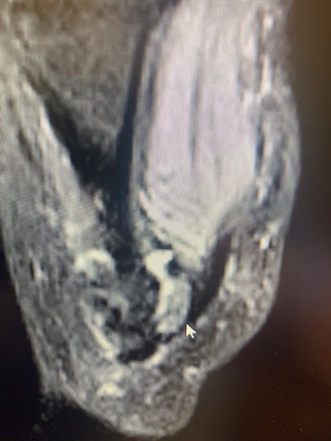
T2 images showing inflamed fibular sesamoid
T2 images showing normal tibial sesamoid


Greeting from San Francisco
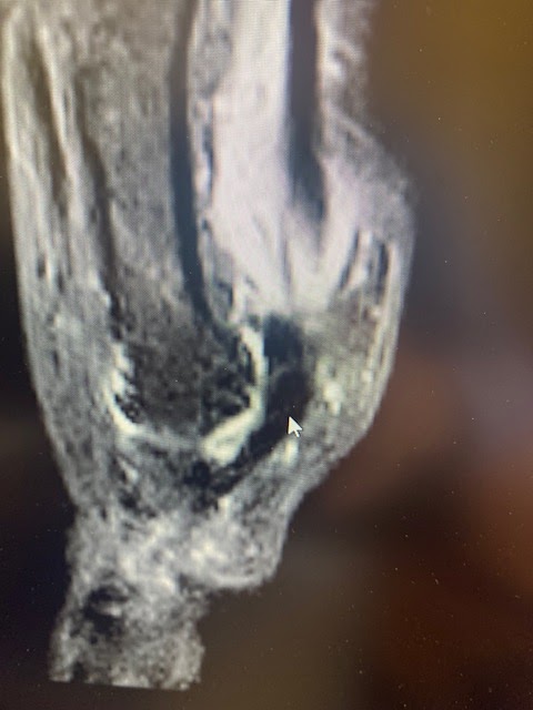
T1 image showing intact stable but injured fibular sesamoid. The grey part of the bone indicates bone activity.
Another T1 image of injured fibular sesamoid. Healing fine. No AVN or avascular necrosis
Great T1 image of both sesamoids. The injured fibular sesamoid has the re-moldeling area in the middle which looks more grey. No AVN or fragmentation.
I have shown both T1 (normal bone white) and T2 (normal bone black) images. When a patient develops AVN, both T1 and T2 are black. Definitely not in this case.
What was your advice to this patient? Surgery?
ReplyDeleteSurgical decision would have to follow failure to progress in treatment. Since, at the time of the images, there was no sign of AVN or fragmentation, conservative treatment seems the best route for now. I would evaluate every 3 months, and typically new MRI every 6 months, if I have gotten the pain consistently to 0-2 VAS, to check progress. Rich
Delete